

Organization of human and mouse skeletal myosin heavy chain gene clusters is highly conserved
- PMID: 10077619
- PMCID: PMC15877
- DOI: 10.1073/pnas.96.6.2958
Organization of human and mouse skeletal myosin heavy chain gene clusters is highly conserved
Abstract
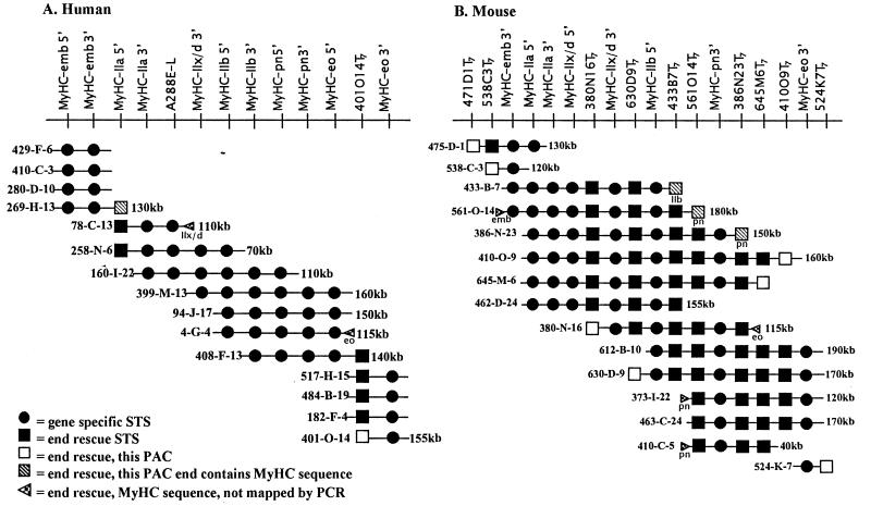
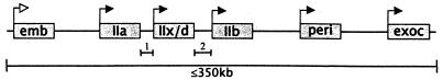
Myosin heavy chains (MyHCs) are highly conserved ubiquitous actin-based motor proteins that drive a wide range of motile processes in eukaryotic cells. MyHC isoforms expressed in skeletal muscles are encoded by a multigene family that is clustered on syntenic regions of human and mouse chromosomes 17 and 11, respectively. In an effort to gain a better understanding of the genomic organization of the skeletal MyHC genes and its effects on the regulation, function, and molecular genetics of this multigene family, we have constructed high-resolution physical maps of both human and mouse loci using PCR-based marker content mapping of P1-artificial chromosome clones. Genes encoding six MyHC isoforms have been mapped with respect to their linear order and transcriptional orientations within a 350-kb region in both human and mouse. These maps reveal that the order, transcriptional orientation, and relative intergenic distances of these genes are remarkably conserved between these species. Unlike many clustered gene families, this order does not reflect the known temporal expression patterns of these genes. However, the conservation of gene organization since the estimated divergence of these species (approximately 75-110 million years ago) suggests that the physical organization of these genes may be significant for their regulation and function.
Figures
References
Publication types
MeSH terms
Substances
Associated data
- Actions
- Actions
- Actions
- Actions
Grants and funding
LinkOut - more resources
Full Text Sources
Other Literature Sources
Molecular Biology Databases
