. 1999 Mar 16;96(6):2982-7.
doi: 10.1073/pnas.96.6.2982.

Vaccination with a recombinant vaccinia virus encoding a "self" antigen induces autoimmune vitiligo and tumor cell destruction in mice: requirement for CD4(+) T lymphocytes

Affiliations

Vaccination with a recombinant vaccinia virus encoding a "self" antigen induces autoimmune vitiligo and tumor cell destruction in mice: requirement for CD4(+) T lymphocytes

W W Overwijk et al. Proc Natl Acad Sci U S A. .

Abstract

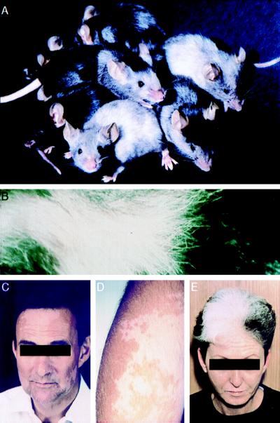
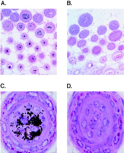
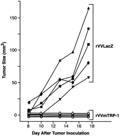
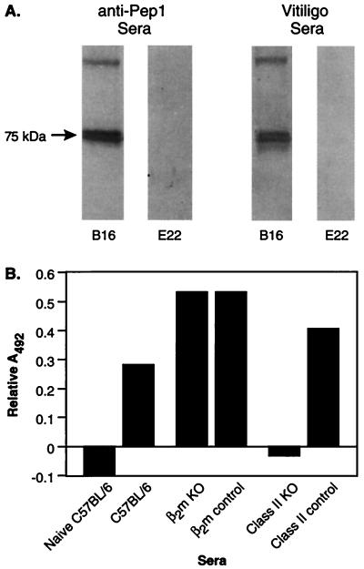
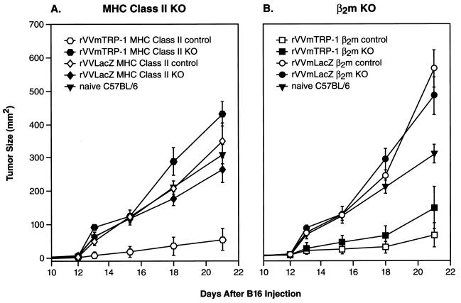
Many human and mouse tumor antigens are normal, nonmutated tissue differentiation antigens. Consequently, immunization with these "self" antigens could induce autoimmunity. When we tried to induce immune responses to five mouse melanocyte differentiation antigens, gp100, MART-1, tyrosinase, and tyrosinase-related proteins (TRP) 1 and TRP-2, we observed striking depigmentation and melanocyte destruction only in the skin of mice inoculated with a vaccinia virus encoding mouse TRP-1. These mice rejected a lethal challenge of B16 melanoma, indicating the immune response against TRP-1 could destroy both normal and malignant melanocytes. Cytotoxic T lymphocytes specific for TRP-1 could not be detected in depigmented mice, but high titers of IgG anti-TRP-1 antibodies were present. Experiments with knockout mice revealed an absolute dependence on major histocompatibility complex class II, but not major histocompatibility complex class I, for the induction of both vitiligo and tumor protection. Together, these results suggest that the deliberate induction of self-reactivity using a recombinant viral vector can lead to tumor destruction, and that in this model, CD4(+) T lymphocytes are an integral part of this process. Vaccine strategies targeting tissue differentiation antigens may be valuable in cancers arising from nonessential cells and organs such as melanocytes, prostate, testis, breast, and ovary.


Figures

Figure 1
Figure 1
Vitiligo-like depigmentation in mice vaccinated twice with rVVmTRP-1. Coat color changes vary in degree, site, and pattern (A). A close-up of depigmented mouse fur is shown (B). These patterns resemble those seen in patients with metastatic malignant melanoma that have been treated with IL-2CE.
Figure 2
Figure 2
Immunization with rVVmTRP-1 induces destruction of cutaneous melanocytes. Mice were vaccinated twice with rVVmTRP-1 (B and D) and developed a patchy and permanent loss of coat color pigmentation. Histologic comparison is with mice receiving control virus rVVLacZ twice (A and C). [Magnifications: ×10 (A and B) and ×100 (C and D).]
Figure 3
Figure 3
Immune responses to the normal melanocyte antigen mTRP-1 can prevent melanoma growth. Mice vaccinated with 2 × 10⁷ plaque-forming units of either rVVLacZ or rVVmTRP-1 and boosted 3 weeks later were challenged with 1 × 10⁵ B16 s.c. after an additional 3 weeks. Mice receiving rVVmTRP-1 developed vitiligo and completely rejected tumor challenge. The experiment was repeated three times with similar results.
Figure 4
Figure 4
Sera from mice immunized with rVVmTRP-1 recognizes mTRP-1 in melanoma cells. (A) Lysates from B16 melanoma (lanes 1 and 3) and E22 thymoma cells (lanes 2 and 4) were subjected to SDS/PAGE, and stained with the mTRP-1 peptide-specific rabbit sera, αPEP1 (lanes 1 and 2), or sera from depigmented mice vaccinated twice with rVVmTRP-1 (lanes 3 and 4). Both sera specifically detected a 75-kDa TRP-1 band in lysate from B16 tumor, but not in lysate from TRP-1 negative E22 control tumor. (B) Induction of mTRP-1-specific antibodies depends on CD4+ T lymphocytes. MHC class II knockout mice, β2m knockout mice, and nontransgenic litter mates were immunized with 2 × 10⁷ plaque-forming units of either rVVLacZ or rVVmTRP-1 and boosted 3 weeks later. Sera from each group of mice were tested by ELISA. Nonspecific absorption of sera from mice immunized twice with rVVLacZ was subtracted. The experiment was repeated twice with similar results.
Figure 5
Figure 5
Autoimmune and antitumor effects of immunization with rVVmTRP-1 are mediated by CD4+ T lymphocytes. β2m knockout mice, MHC class II knockout mice, or respective nontransgenic litter mates were immunized with 2 × 10⁷ plaque-forming units of either rVVLacZ or rVVmTRP-1, boosted 3 weeks later and challenged with 1 × 10⁵ B16 s.c. after an additional 3 weeks. Both β2m knockout mice and nontransgenic litter mates receiving rVVmTRP-1 developed vitiligo and were protected against B16 tumor challenge (B). In the same experiment, MHC class II knockout mice never developed vitiligo after immunization with rVVmTRP-1, nor were they protected against B16 tumor challenge (A). Nontransgenic litter mates receiving rVVmTRP-1 developed vitiligo and were protected against B16 tumor challenge, indicating a critical role for CD4+ T lymphocytes in the induction of vitiligo and tumor protection. The experiment was repeated twice with similar results.

References

    1. Prehn R T, Main J M. J Natl Cancer Inst. 1957;18:769–774. - PubMed
    1. Barth R J J, Bock S N, Mule J J, Rosenberg S A. J Immunol. 1990;144:1531–1537. - PubMed
    1. Melief C J, Kast W M. Immunol Rev. 1995;145:167–177. - PubMed
    1. Overwijk W W, Tsung A, Irvine K R, Parkhurst M R, Goletz T J, Tsung K, Carroll M W, Liu C, Moss B, Rosenberg S A, Restifo N P. J Exp Med. 1998;188:277–286. - PMC - PubMed
    1. Boon T, Coulie P G, van den Eynde B. Immunol Today. 1997;18:267–268. - PubMed

MeSH terms