Comparative Study
. 1999 Mar 16;96(6):3029-34.
doi: 10.1073/pnas.96.6.3029.

In vitro comparison of the antigen-binding and stability properties of the various molecular forms of IgA antibodies assembled and produced in CHO cells

Affiliations
Comparative Study

In vitro comparison of the antigen-binding and stability properties of the various molecular forms of IgA antibodies assembled and produced in CHO cells

J Berdoz et al. Proc Natl Acad Sci U S A. .

Abstract

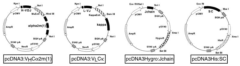
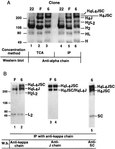
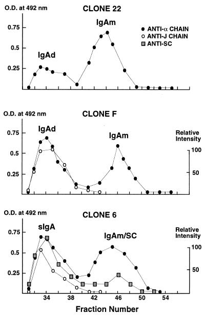
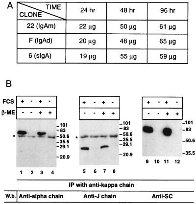
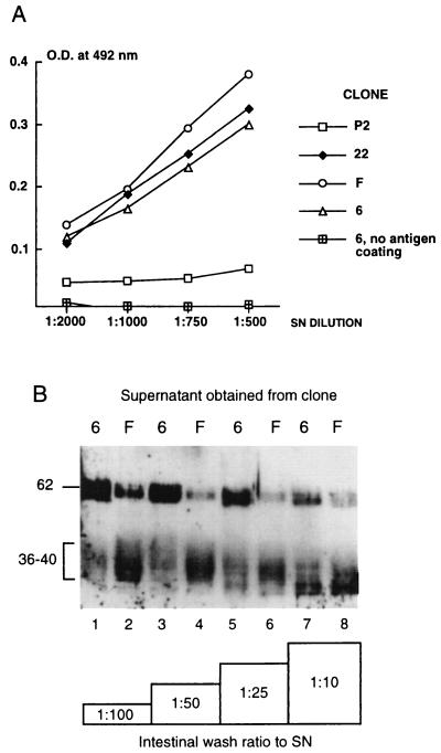
The hallmark of a mucosal immune response is the production of antigen-specific secretory IgA (S-IgA) antibodies in external secretions. S-IgA consists of ten polypeptides produced in two different cell lineages. The heavy and light chains in plasma cells assemble into IgA, which on association with J chain become polymerized, whereas secretory component (SC) is added during transport across the epithelium. Recombinant chimeric mouse-human monomeric, dimeric, and S-IgA antibodies have been produced in a single CHO cell sequentially transfected with expression vectors carrying three independent selective markers for chimeric heavy and light chains, human J chain, and human SC, respectively. Biochemical characterization of the various molecular forms indicates that the assembly of the various polypeptides resulted in species of the expected size and covalence. All chimeric IgA antibodies retained the antigen-binding capacity of the parent mouse IgA antibody. The resistance of S-IgA to protease-rich intestinal washes was enhanced when compared with dimeric IgA lacking associated SC. Up to 20 micrograms of recombinant S-IgA per 1 x 10(6) cells were recovered in 24 h with the best producing clones. We conclude that CHO cells programmed de novo with four different genetic elements can assemble functional chimeric S-IgA.


Figures

Figure 1
Figure 1
Schematic representation of the expression vectors used in this study. All constructs were derived from pcDNA3. Plasmids pcDNA3:VHCα2m(1) and pcDNA3:VLCκ contain the original ORF for neomycin resistance (neoR), pcDNA3Hygro:Jchain the ORF for hygromycin B resistance (hygroR), and pcDNA3His:SC the ORF for histidinol resistance (hisR). The two black boxes called H-VDJ and L-VJ represent the genomic DNA coding for the murine heavy and light variable regions isolated from HNK-20 hybridoma. The other black boxes contain the human genomic region comprising the three α-chain domains, the human genomic region coding for the κ-chain, and the human cDNAs for the J chain and SC, respectively. Striped segments span the μ and κ enhancers obtained from mouse genomic fragments. The restriction sites used for the construction of the vectors are indicated.
Figure 2
Figure 2
Polymeric IgA molecules assemble in CHO cells. (A) Cell culture SN from clones 22, F, and 6 were concentrated by precipitation with TCA, or immunoprecipitation (IP) with anti-κ-chain antiserum/protein A Sepharose beads. After separation onto a 5–13% polyacrylamide gradient gel in SDS under nonreducing conditions, the proteins were transferred onto membranes and detected by using an anti-α-chain antiserum. Molecular weight markers run in parallel served to identify the various polymeric forms marked as combination of H (heavy), L (light), and J (J) chains and SC (SC). (B) Cell culture SN from clones F and 6 were concentrated with anti-κ-chain antiserum/protein A Sepharose beads and processed as in A. Blotted proteins were incubated with antisera directed against the κ-chain (lanes 1 and 2), the J chain (lanes 3 and 4), and SC (lane 5) to confirm the nature of species identified in A. Note that each specific antiserum has a distinct sensitivity, which explains the different relative intensity of the multiple molecular forms.
Figure 3
Figure 3
Analysis of the composition of IgA forms secreted by clones 22, F, and 6. Two hundred microliters of crude SN was separated by chromatography on a Superose 12 column (Pharmacia) equilibrated and run in PBS. Fractions of 250 μl were collected and analyzed by ELISA (α-chain and SC) or Western blot (J chain). For ELISA, the resulting OD at 492 nm is plotted. For Western blot analysis, half of the fraction was concentrated by TCA precipitation before loading on a 15% polyacrylamide/SDS gel. After transfer, the protein was detected with rabbit anti-J chain antiserum, followed by mouse anti-rabbit antibody conjugated with HRP and chemiluminescence. Films were scanned by using an Elscript 400-AT/SM densitometer, and values were plotted with the strongest intensity arbitrarily fixed as 100%.
Figure 4
Figure 4
Effect of time, FCS, and 2-mercaptoethanol on IgA expression. (A) Level of IgA production in SN of clones 22, F, and 6, expressed in μg per 1 × 10⁶ cells as a function of time. IgA were measured by ELISA by using purified dIgA (clones 22 and F) and S-IgA (clone 6) as standards. (B) Effect of FCS and 2-mercaptoethanol (β-ME) on expression of S-IgA by clone 6. The cell culture SN was concentrated by immunoprecipitation (IP) with anti-κ-chain antiserum/protein A Sepharose beads, and separated by SDS/PAGE under reducing conditions. After transfer onto membranes, polypeptides were detected with antisera to the α-chain, the J chain, and SC. Molecular weight standards are indicated on the side of the gel. Asterisks indicate cross-reactive bands corresponding to the IgG heavy chain in the antiserum used for immunoprecipitation.
Figure 5
Figure 5
Antigen-binding activity and comparative digestion patterns of IgA molecular forms secreted by CHO cells. (A) Crude CHO SN were incubated in wells coated with RSV whole lysate containing the glycoprotein F antigen. The bound antibodies were detected with biotinylated antibodies to the α-chain, followed by streptavidin conjugated with HRP. The initial concentration of each antibody solution obtained from clones 22, F, and 6 was standardized to 20 μg/ml by diluting the SN with cell culture medium. Results are means of three separate experiments done in duplicates. SN of untransfected CHO cell clone P2 and wells noncoated with the antigen were used as controls. Lower dilutions of the SNs resulted in loss of linearity of the response curve. (B) FCS-free SN containing identical amounts of recombinant IgA were combined with four different dilutions of pooled intestinal washes, resulting in an antibody/intestinal wash ratio ranging from 1:100 to 1:10. Digestion was carried out for 16 h at 37°C. Degradation products were separated by SDS/PAGE under reducing conditions and assessed by immunodetection of the α-chain. The position of the intact α-chain lies at 62 kDa, whereas degradation produces a series of bands at 36–40 kDa (14).

References

    1. Neutra M R. Curr Top Microbiol Immunol. 1999;236:17–32. - PubMed
    1. Corthésy B, Kraehenbuhl J-P. Curr Top Microbiol Immunol. 1999;236:93–111. - PubMed
    1. Mostov K E. Annu Rev Immunol. 1994;12:63–84. - PubMed
    1. Mazanec M B, Nedrud J G, Kaetzel C S, Lamm M E. Immunol Today. 1993;14:430–435. - PubMed
    1. Michetti P, Mahan M J, Slauch J M, Mekalanos J J, Neutra M R. Infect Immun. 1992;60:1786–1792. - PMC - PubMed

Publication types

Substances

LinkOut - more resources