. 1999 Mar 16;96(6):3081-6.
doi: 10.1073/pnas.96.6.3081.

Epidermal growth factor system regulates mucin production in airways

Affiliations

Epidermal growth factor system regulates mucin production in airways

K Takeyama et al. Proc Natl Acad Sci U S A. .

Abstract

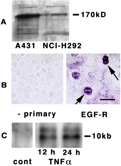
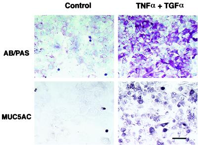
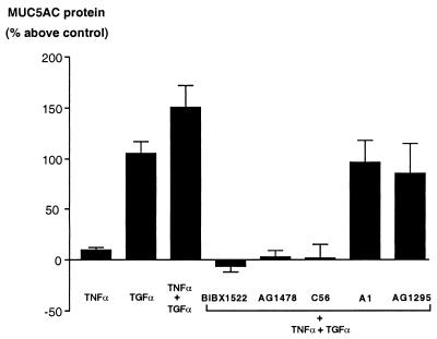
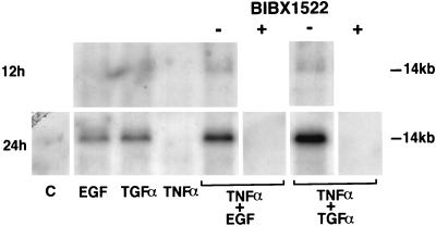
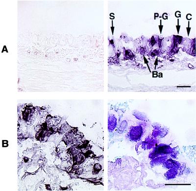
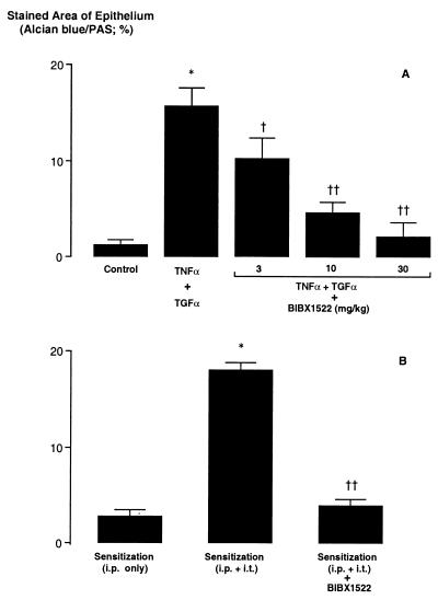
Goblet-cell hyperplasia is a critical pathological feature in hypersecretory diseases of airways. However, the underlying mechanisms are unknown, and no effective therapy exists. Here we show that stimulation of epidermal growth factor receptors (EGF-R) by its ligands, EGF and transforming growth factor alpha (TGFalpha), causes MUC5AC expression in airway epithelial cells both in in vitro and in vivo. We found that a MUC5AC-inducing epithelial cell line, NCI-H292, expresses EGF-R constitutively; EGF-R gene expression was stimulated further by tumor necrosis factor alpha (TNFalpha). EGF-R ligands increased the expression of MUC5AC at both gene and protein levels, and this effect was potentiated by TNFalpha. Selective EGF-R tyrosine kinase inhibitors blocked MUC5AC expression induced by EGF-R ligands. Pathogen-free rats expressed little EGF-R protein in airway epithelial cells; intratracheal instillation of TNFalpha induced EGF-R in airway epithelial cells, and subsequent instillation of EGF-R ligands increased the number of goblet cells, Alcian blue-periodic acid-Schiff staining (reflecting mucous glycoconjugates), and MUC5AC gene expression, whereas TNFalpha, EGF, or TGFalpha alone was without effect. In sensitized rats, three intratracheal instillations of ovalbumin resulted in EGF-R expression and goblet-cell production in airway epithelium. Pretreatment with EGF-R tyrosine kinase inhibitor, BIBX1522, prevented goblet-cell production both in rats stimulated by TNFalpha-EGF-R ligands and in an asthma model. These findings suggest potential roles for inhibitors of the EGF-R cascade in hypersecretory diseases of airways.


Figures

Figure 1
Figure 1
Expression of EGF-R in cultured cells. (A) Immunoblotting of EGF-R in A431 and in NCI-H292 cells. Cells were examined after becoming confluent. Lysates were electrophoresed in 8% acrylamide gels and blotted with anti-EGF-R antibody. Molecular mass of marker protein is reported on the right. (B) Immunocytochemical analysis with anti-EGF-R antibody in cultures of NCI-H292 cells. At confluence, positive staining was seen in most of cells, and certain cells had more intense staining (arrows, right side). In the absence of the primary antibody, no staining was seen (left side). (Bar = 25 μm.) (C) Northern analysis of EGF-R in NCI-H292 cells was performed on total RNA extracted from confluent cultures incubated with TNFα (20 ng/ml) for 12 or 24 h. The RNA (10 μg) was electrophoresed on a formaldehyde-agarose gel, transferred to a nylon membrane, and hybridized with the 32P-labeled EGF-R cDNA probe. After hybridization, the membrane was washed and autoradiographed.
Figure 2
Figure 2
AB–PAS staining of NCI-H292 cells for identification of mucous glycoconjugates (Upper) and immunocytochemical analysis for MUC5AC protein (Lower). Immunocytochemical analysis with an anti-MUC5AC antibody showed positive staining in only a few cells in the control state in a similar pattern to that of AB–PAS staining. However, incubation with TNFα (20 ng/ml) plus TGFα (25 ng/ml) increased both the PAS-positive staining and MUC5AC protein markedly. (Bar = 50 μm.)
Figure 3
Figure 3
ELISA for MUC5AC in NCI-H292 cells. MUC5AC protein was measured as described in Methods. TNFα caused little expression of MUC5AC protein (8.7 ± 3.8%, n = 3); TGFα alone caused ≈100% increase in MUC5AC protein, an effect that was potentiated by coincubation with TNFα (149.5 ± 21.7%, n = 6). The stimulatory effect of TNFα plus TGFα was inhibited by each of the three selective EGF-R tyrosine kinase inhibitors (BIBX1522, −6.0 ± 5.5%; tyrphostin AG1478, 2.1 ± 6.7%; Compound 56, 1.0 ± 14.5%; n = 3), whereas a negative control for tyrphostins (tyrphostin A1) and a selective inhibitor of platelet-derived growth factor (tyrphostin AG1295) were without effect.
Figure 4
Figure 4
Northern analysis for MUC5AC gene expression in NCI-H292 cells. Total RNA was extracted from the cells, and 10 μg of RNA was electrophoresed on a formaldehyde-agarose gel, transferred to a nylon membrane, and hybridized with the 32P-labeled MUC5AC cDNA probe. After hybridization, the membrane was washed and autoradiographed. Cultures were obtained with medium alone (C), EGF or TGFα (25 ng/ml), TNFα (20 ng/ml), or the combination of TNFα plus either EGF or TGFα for 12 h (Upper) or 24 h (Lower) on MUC5AC gene expression. Cultures were also obtained with TNFα plus either EGF or TGFα after preincubation with EGF-R tyrosine kinase inhibitor (BIBX1522; 10 μg/ml; Lower); the inhibitor prevented MUC5AC gene expression. Three different experiments showed similar results.
Figure 5
Figure 5
Immunohistochemical analysis of EGF-R with an anti-EGF-R antibody in pathogen-free rats. (A) TNFα-treated rats. Control animals showed little EGF-R staining (Left); 24 h after intratracheal instillation of TNFα (200 ng, 100 μl), EGF-R-positive staining was present in goblet cells (G), pre-goblet cells (P-G), nongranulated secretory cells (S), and basal cells (Ba), but not in ciliated cells. (Bar = 50 μm.) (B) Ovalbumin sensitization. After three intratracheal instillations of ovalbumin (0.1%, 100 μl), EGF-R immunoreactivity was strongly expressed in goblet and pre-goblet cells (Left), the same cells that stained positively with AB–PAS (Right). (Bar = 50 μm.)
Figure 6
Figure 6
Effect of EGF-R tyrosine kinase inhibitor (BIBX1522) on production of goblet cells (expressed as % stained area of airway epithelium occupied by AB–PAS-positive stained cells). (A) Stimulation with TNFα (200 ng, 100 μl). Tracheal instillation of TNFα, followed by the EGF-R ligand TGFα, increased goblet-cell production significantly (n = 5; ∗, P < 0.0001), an effect that was inhibited by pretreatment with BIBX1522 (3–30 mg/kg, i.p.) dose-dependently (n = 5; P compared with TNFα followed by TGFα: , P = 0.003; ††, P < 0.0001). (B) Ovalbumin sensitization. Animals given ovalbumin intraperitoneally (i.p.) only showed little AB–PAS-positive staining in bronchial epithelium. Animals first sensitized with ovalbumin (OVA) i.p., followed by three intratracheal (i.t.) instillations of OVA, showed a marked increase in AB–PAS-positive staining (n = 5; ∗, P < 0.0001). Pretreatment with BIBX1522 (10 mg/kg, i.p.) inhibited OVA-induced production of goblet cells (n = 5; ††, P < 0.0001).

References

    1. Snider G L, Faling L J, Rennard S I. In: Textbook of Respiratory Medicine. Murray J F, Nadel J A, editors. New York: Saunders; 1994. , Chap. 41.
    1. Boat T F, Boucher R C. In: Textbook of Respiratory Medicine. Murray J F, Nadel J A, editors. New York: Saunders; 1994. , Chapter 43.
    1. Fahy J V, Schuster A, Ueki I, Boushey H A, Nadel J A. Am Rev Respir Dis. 1992;146:1430–1433. - PubMed
    1. Cardell B S, Pearson R S B. Thorax. 1959;14:341–352.
    1. Dunnill M S. J Clin Pathol. 1960;13:27–33. - PMC - PubMed

Publication types