. 1999 Mar 16;96(6):3188-93.
doi: 10.1073/pnas.96.6.3188.

The role of an alternative sigma factor in motility and pilus formation in the cyanobacterium Synechocystis sp. strain PCC6803

Affiliations

The role of an alternative sigma factor in motility and pilus formation in the cyanobacterium Synechocystis sp. strain PCC6803

D Bhaya et al. Proc Natl Acad Sci U S A. .

Abstract

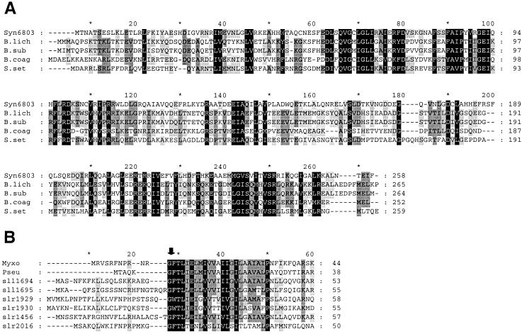
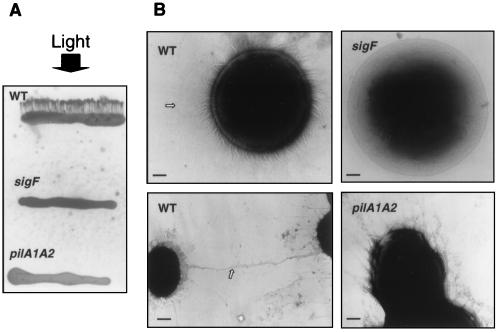
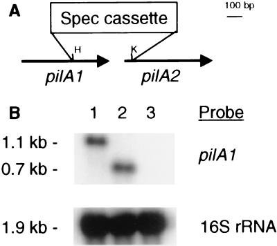
Disruption of a gene for an alternative sigma factor, designated sigF, in the freshwater, unicellular cyanobacterium Synechocystis sp. strain PCC6803 resulted in a pleiotropic phenotype. Most notably, this mutant lost phototactic movement with a concomitant loss of pili, which are abundant on the surface of wild-type cells. The sigF mutant also secreted both high levels of yellow-brown and UV-absorbing pigments and a polypeptide that is similar to a large family of extracellular proteins that includes the hemolysins. Furthermore, the sigF mutant had a dramatically reduced level of the transcript from two tandemly arranged pilA genes (sll1694 and sll1695), which encode major structural components of type IV pili. Inactivation of these pilA genes eliminated phototactic movement, though some pili were still present in this strain. Together, these results demonstrate that SigF plays a critical role in motility via the control of pili formation and is also likely to regulate other features of the cell surface. Furthermore, the data provide evidence that type IV pili are required for phototactic movement in certain cyanobacteria and suggest that different populations of pili present on the Synechocystis cell surface may perform different functions.


Figures

Figure 1
Figure 1
(A) Alignment of SigF of Synechocystis (Syn6803) with SigB of Bacillus licheniformis (B.lich) and B. subtilis (B.sub), SigF of Bacillus coagulans (B.coag), and a sigma factor from Streptomyces setonii (S.set). Black boxes indicate identical or conserved residues at the same position in all five sequences while dark gray and light gray boxes indicate identical or conserved residues in four of the five and three of the five sequences, respectively. The GenBank accession nos. for the B. licheniformis, B. subtilis, B. cogulans and S. setonii sequences are AF034567, M14508, Z44161, and D17466, respectively. (B) Alignment of the conserved N-termini of six putative PilA-like sequences (encoded by sll1694, sll1695, slr1929, slr1930, slr1456, and slr2016) from Synechocystis with PilA polypeptides of M. xanthus (Myxo) and P. aeruginosa (Pseu). The GenBank accession nos. for the M. xanthus and P. aeruginosa pilA genes are L39904 and L37109, respectively. The arrow indicates the putative cleavage site of the precursor with phenylalanine (F) being the first amino acid of the mature protein. Only the leader sequence and the highly conserved 32 aa at the amino terminus of the mature protein are shown. Black boxes indicate identical or conserved residues at the same position in all eight sequences, while dark gray and light gray boxes indicate identical or conserved residues in at least six of eight and five of eight sequences, respectively. Comparisons were performed by using clustal or the gapped blast search available through the National Center for Biotechnology Information (http://www.ncbi.nlm.nih.gov/). Alignments were plotted by using the genedoc program.
Figure 2
Figure 2
(A) Spectra of wild-type (WT) and sigB cultures. Spectra (250–800 nm) were measured from 6-day-old cultures of the sigF mutant (a) and WT (b), the cell pellet of the sigF mutant (c) and WT (d), and the growth media of the sigF mutant (e) and WT (f). Spectra are offset along the ordinate and normalized to cell density as reflected by absorbance at 750 nm. (B) SDS/PAGE of polypeptides precipitated from the growth media from WT (lane 2) and sigF mutant (lane 3) cultures and stained with Coomassie blue. Molecular mass markers (in kDa) are shown in lane 1.
Figure 3
Figure 3
(A) Directional motility assay. Five to 10 microliters of logarithmically growing wild-type cells (WT) and the sigF and pilA1A2 mutants were streaked as an approximately 1-mm thick line onto solid (0.4% agar) BG-11 medium containing 15 mM glucose. The cells were subjected to unidirectional light (indicated by an arrow) of 40 μmol photons·m−2⋅s−1 for 48 h. The temperature during the incubation was 30°C. Note the finger-like projections emerging from the WT streak. (B) Transmission electron microscopy of whole cells. Logarithmically growing WT (Left), sigF mutant (Upper Right) and pilA1A2 mutant (Lower Right) cells. The cells were negatively stained with 1% phosphotungstic acid and examined by using a Phillips CM12 microscope. The arrow in WT (Upper Left) points to a very long pilus while the arrow in WT (Lower Left) points to pili that appear to connect neighboring cells. The bars represent 0.28 μm (Upper Left), 0.24 μm (Upper Right), 0.74 μm (Lower Left), and 0.31 μm (Lower Right).
Figure 4
Figure 4
(A) The tandemly arranged pilA1 (sll1694) and pilA2 (sll1695) genes showing insertion of the spectinomycin cassette into the region between the HpaI (H) and KpnI (K) sites. (B) Hybridization of RNA from wild-type cells (lane 1) and the pilA1A2 (lane 2) and sigF (lane 3) mutants to probes for the pilA1 gene (Upper) or 16S rRNA (Lower), which was used as a loading control.

References

    1. Häder D-P. In: The Cyanobacteria. Fay P, Baalen C V, editors. Amsterdam: Elsevier; 1987. pp. 325–346.
    1. Castenholz R W. In: The Biology of Cyanobacteria. Carr N G, Whitton B A, editors. Botanical Monographs 19. Berkeley: Univ. of California Press; 1982. pp. 413–440.
    1. Hoiczyk E, Baumeister W. J Bacteriol. 1995;177:2387–2395. - PMC - PubMed
    1. Hoiczyk E, Baumeister W. Mol Microbiol. 1997;26:699–708. - PubMed
    1. Waterbury J B, Willey J M, Franks D G, Valois F W, Watson S W. Science. 1985;230:74–75. - PubMed

Publication types

LinkOut - more resources