. 1999 Mar 16;96(6):3239-44.
doi: 10.1073/pnas.96.6.3239.

Calcium/calmodulin-dependent protein kinase II is associated with the N-methyl-D-aspartate receptor

Affiliations

Calcium/calmodulin-dependent protein kinase II is associated with the N-methyl-D-aspartate receptor

A S Leonard et al. Proc Natl Acad Sci U S A. .

Abstract

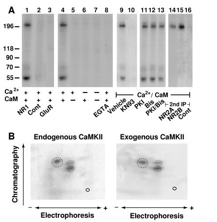
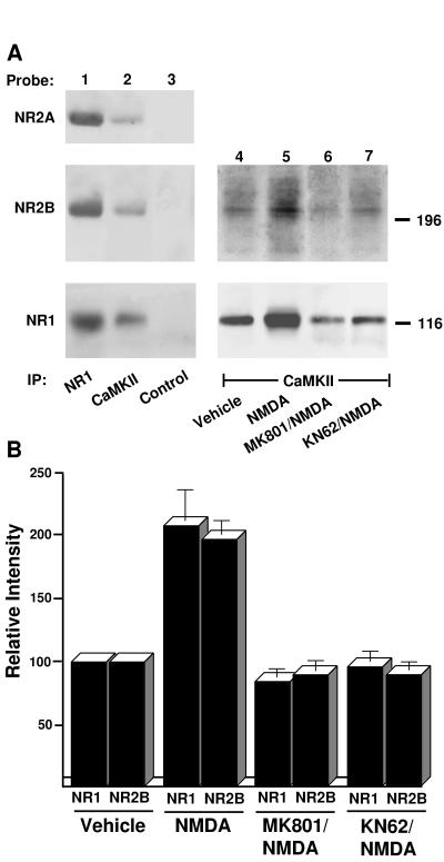
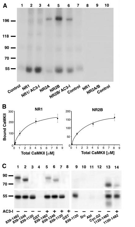
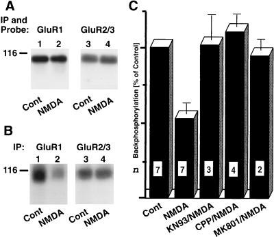
The molecular basis of long-term potentiation (LTP), a long-lasting change in synaptic transmission, is of fundamental interest because of its implication in learning. Usually LTP depends on Ca2+ influx through postsynaptic N-methyl-D-aspartate (NMDA)-type glutamate receptors and subsequent activation of Ca2+/calmodulin-dependent protein kinase II (CaMKII). For a molecular understanding of LTP it is crucial to know how CaMKII is localized to its postsynaptic targets because protein kinases often are targeted to their substrates by adapter proteins. Here we show that CaMKII directly binds to the NMDA receptor subunits NR1 and NR2B. Moreover, activation of CaMKIIalpha by stimulation of NMDA receptors in forebrain slices increase this association. This interaction places CaMKII not only proximal to a major source of Ca2+ influx but also close to alpha-amino-3-hydroxy-5-methylisoxazole-4-propionic acid (AMPA)-type glutamate receptors, which become phosphorylated upon stimulation of NMDA receptors in these forebrain slices. Identification of the postsynaptic adapter for CaMKII fills a critical gap in the understanding of LTP because CaMKII-mediated phosphorylation of AMPA receptors is an important step during LTP.


Figures

Figure 1
Figure 1
CaMKII activity is associated with NMDA receptors. (A) NMDA receptor complexes were extracted with 1% deoxycholate from crude rat forebrain membrane fractions, and immunoprecipitations were performed with the NR1-specific antibody αNR1 (lanes 1 and 4–16), control mouse IgG (lane 2), or αGluR1 for precipitation of AMPA receptors (lane 3). Immunocomplexes were incubated in phosphorylation buffer containing Ca2+, calmodulin, EGTA (1 mM), KN93 (50 μM), PKI (1 μM), or the bisindolylmaleimide GF109203X (Bis; 1 μM) as indicated. Samples were either subjected directly to SDS/PAGE (lanes 1–13) or dissociated with SDS before a second immunoprecipitation with the antibodies specific for NR2A or 2B or with control rabbit antibodies was performed (lanes 14–16). Phosphorylated polypeptides were detected by autoradiography. Positions of marker proteins are indicated on the left (in kDa). Similar results were obtained in three different experiments. (B) For phosphopeptide mapping of the NR2B subunit phosphorylated by endogenous kinase (Left), NMDA receptors were solubilized, phosphorylated, and dissociated with SDS before reprecipitation of NR2B and SDS/PAGE. To obtain phosphopeptide maps of NR2B phosphorylated by exogenous CaMKII (Right), crude rat forebrain membrane fractions were extracted with SDS. NR2B was immunoprecipitated and phosphorylated with recombinant CaMKII before SDS/PAGE. Of note, no endogenous kinase activity was detectable when NR2B was immunoprecipitated after dissociation with SDS (see Fig. 3A, lane 9). Tryptic two-dimensional phosphopeptide maps were produced in three independent experiments. Main phosphopeptides are circled.
Figure 2
Figure 2
Coimmunoprecipitation of NMDA receptors with CaMKII. (A) To test whether NMDA receptors are associated with CaMKII in vivo, crude membrane fractions were prepared from total rat forebrain and NMDA receptors were solubilized with deoxycholate. After immunoprecipitation with antibodies against NR1, a mixture of the antibodies against CaMKIIα and β (19) or with control mouse IgG, immunoblotting was performed (lanes 1–3) with antibodies against NR2A (Left Top), NR2B (Left Middle), and NR1 (Left Bottom). To measure the effect of NMDA receptor-mediated Ca2+ influx on CaMKII association with NMDA receptors in intact neurons, acute cortical slices were prepared and treated under control conditions (vehicle) or with 200 μM NMDA for 5 min in the absence or presence of 50 μM MK801 or 50 μM KN62. Crude membrane fractions were prepared and solubilized with deoxycholate. Immunoprecipitations were performed with a mixture of the antibodies against CaMKIIα and β (19) before immunoblotting with αNR1 (Right Lower) or a mixture of antibodies against NR2A and NR2B (Right Upper) (lanes 4–7). Similar results were obtained in two other experiments; the results of all three cortical slice experiments were quantified by densitometry of the immunoblotting signals (33) and are summarized in B. Bars = SEM.
Figure 3
Figure 3
CaMKIIα binding to NR1 and NR2B. (A and B) Crude membrane fractions were extracted and NMDA receptor subunits were dissociated with SDS before immunoprecipitation of individual subunits as indicated on the bottom (A) or top (B). Control immunoprecipitations were performed with nonspecific murine (A, lane 1) or rabbit IgG (A, lanes 7 and 10). Recombinant CaMKIIα was preincubated under phosphorylation conditions in the presence of [γ-32P]ATP for labeling by autophosphorylation, and immunocomplexes were incubated with the whole phosphorylation mixture. The signal at 55 kDa (A, lanes 2, 3, 5, and 6) reflects binding of recombinant and autophosphorylated CaMKIIα rather than the presence of the endogenous kinase because no phosphorylation was detectable if recombinant CaMKIIα was omitted (A, lanes 8–10). Phosphorylation of NR2B (A, lane 6) but not association of CaMKIIα with NR1 (A, lane 3) or NR2B (A, lane 6) was inhibited if the competitive catalytic site inhibitor AC3-I (20 μM) was added after autophosphorylation of CaMKIIα (27). Similar results were obtained in two other experiments. Binding of CaMKIIα to NR1 and NR2B was saturable (B; the amounts of CaMKIIα associated with NR1 or NR2B were quantified after SDS/PAGE by using PhosphorImager analysis; values are means ± SEM; n = 3). (C) CaMKIIα was preincubated under phosphorylation conditions with [γ-32P]ATP for labeling by autophosphorylation. Twenty microliters of glutathione Sepharose loaded with approximately 2 μg (as confirmed by staining with Coomassie brilliant blue) of GST or GST fusion proteins (as indicated at the bottom) then was added (together with 20 μM AC3-I when indicated) before samples were washed and analyzed by SDS/PAGE and autoradiog raphy. GST-NR2B839–1482, 839–1346, and 1120–1482, but not 839–1120 or GST alone, were phosphorylated by CaMKII in an AC3-I- sensitive manner (Upper). Autophosphorylated CaMKIIα bound equally well to all four GST-NR2B fusion proteins but not to GST alone or to nonrelevant GST fusion proteins with the Src or Abl SH3 domain or cyclin G2 (Lower).
Figure 4
Figure 4
NMDA receptor activation stimulates phosphorylation of GluR1 by CaMKII. Cortical slices were pretreated with the bisindolylmaleimide GF109203X for 20 min to inhibit PKC-mediated phosphorylation and incubated for 5 min with or without NMDA (200 μM) as indicated (22, 33). After solubilization with Triton X-100, AMPA receptor complexes were immunoprecipitated with a mixture of antibodies against GluR1 and GluR2/3. (A) After dissociated of the immunocomplexes with SDS and reprecipitated with either αGluR1 (lanes 1 and 2) or αGluR2/3 (lanes 3 and 4), immunoblotting was performed with the corresponding antibodies to ensure that equal amounts of GluR1 and GluR2/3 were present in extracts from control and NMDA-treated slices. (B and C) AMPA receptor complexes were back-phosphorylated with recombinant CaMKIIα, dissociated with SDS, and reprecipitated with either GluR1- or GluR2/3-specific antibodies (lanes 1 and 2 and 3 and 4, respectively). After SDS/PAGE, samples were analyzed by autoradiography (B) or quantified by PhosphorImager analysis (C). Some slices were preincubated with 50 μM CPP, MK801, or KN93 before NMDA application. Values are means ± SEM; n is indicated in each column.

References

    1. Braun A P, Schulman H. Annu Rev Physiol. 1995;57:417–445. - PubMed
    1. Ghosh A, Greenberg M E. Science. 1995;268:239–247. - PubMed
    1. Petralia R S, Wenthold R J. J Comp Neurol. 1992;318:329–354. - PubMed
    1. Petralia R S, Yokotani N, Wenthold R J. J Neurosci. 1994;14:667–696. - PMC - PubMed
    1. Bliss T V, Collingridge G L. Nature (London) 1993;361:31–39. - PubMed

Publication types

MeSH terms

Substances

LinkOut - more resources