. 1999 Mar 16;96(6):3245-50.
doi: 10.1073/pnas.96.6.3245.

Stimulated changes in localized cerebral energy consumption under anesthesia

Affiliations

Stimulated changes in localized cerebral energy consumption under anesthesia

R G Shulman et al. Proc Natl Acad Sci U S A. .

Abstract

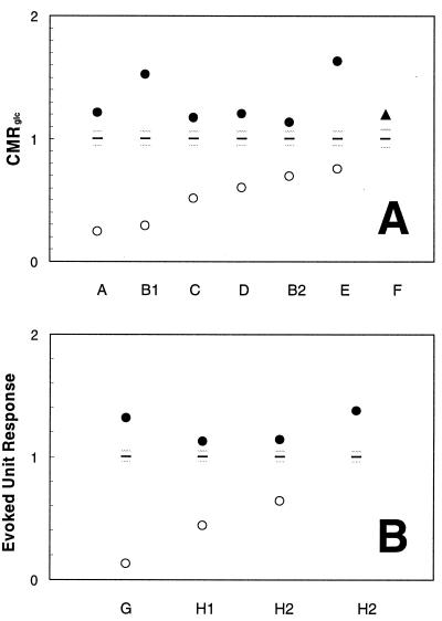
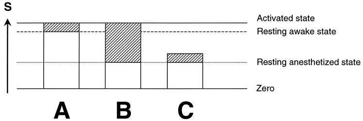
Focal changes in the cerebral metabolic rate of glucose utilization (CMRglc) are small (10-40%) during sensory activation in awake humans, as well as in awake rodents and primates (20-50%). They are significantly larger (50-250%) in sensory activation studies of anesthetized rats and cats. Our data, in agreement with literature values, show that in the resting anesthetized state values of CMRglc are lower than in the resting nonanesthetized state whereas the final state values, reached upon activation, are similar for the anesthetized and nonanesthetized animals. The lower resting anesthetized state values of CMRglc explain why the increments upon activation from anesthesia are larger than when starting from the nonanesthetized conditions. Recent 13C NMR measurements in our laboratory have established a quantitative relationship between the energetics of glucose oxidation, CMRglc (oxidative), and the flux of the glutamate/gamma-aminobutyric acid/glutamine neurotransmitter cycle, Vcycle. In both the resting awake value of CMRglc(oxidative), and its increment upon stimulation, a large majority (approximately 80%) of the brain energy consumption is devoted to Vcycle. In the differencing methods of functional imaging, it is assumed that the incremental change in the measured signal represents the modular activity that supports the functional response. However, the same amount of activity must be present during the response to stimulation, irrespective of the initial basal state of the cortex. Thus, whereas the incremental signals of DeltaCMRglc can localize neurotransmitter activity, the magnitude of such activity during the response is represented by the total localized CMRglc, not the increment.


Figures

Figure 1
Figure 1
Data from Tables 1 and 2, respectively, were used to normalize the resting anesthetized state and activated state values of CMRglc (A) and evoked unit response (B) from rats, cats, and monkeys on the same vertical scale. The mean ± SD values of each resting awake state depicted possible variations in the awake state (black horizontal line represents mean and the gray horizontal lines represent the SD). The activated state reached from the resting anesthetized state is higher than the resting awake state, explaining why the increments upon activation from anesthesia are larger than when starting from the awake resting state. For the anesthetized animal the increment with stimulation raises the local activity to a level that is similar to that reached upon stimulation of the nonanesthetized awake state, which supports the suggestion that a particular magnitude of activity is a required response to a sensory stimulus and not a particular increment. Refer to Tables 1 and 2 for references.
Figure 2
Figure 2
Schematic representation of possible increase in activity upon stimulation for an animal that is nonanesthetized (A) and anesthetized (B and C). The incremental functional imaging signal, ΔS, obtained by differencing, is represented by the shaded rectangles and is normally used to reveal the focally activated regions. The remaining neuronal activity, which is represented by white rectangles, is ignored by the differencing method. For the anesthetized animal, the incremental activity shown by the shaded areas would be larger for B than for C. If the incremental activity needed to perform the task were modular, and therefore independent of the initial activity, then the increment should be the same for the resting anesthetized or resting awake states (compare A and C). If, on the other hand, the final level rather than the increment was needed to support the activity upon responding to stimulation, the incremental signal from anesthesia would be much larger than in the response from the awake state. The data summarized in Tables 1 and 2 and Fig. 1 show that the incremental signal under anesthesia is actually larger, and that during stimulation, S always rises to approximately the same absolute level independent of the initial basal state. These results support a view in which a particular magnitude of neuronal activity is required for a task (B), not a particular increment (C).

References

    1. Roy C S, Sherrington C S. J Physiol (London) 1890;11:85–108. - PMC - PubMed
    1. Ritchie J M. J Physiol. 1967;188:309–329. - PMC - PubMed
    1. Creutzfeldt O D. In: Brain Work: The Coupling of Function Metabolism and Blood Flow in the Brain. Ingvar D H, Lassen N A, editors. Munksgaard, Copenhagen: Alfred Benzon Foundation; 1975. , Alfred Benzon Symposium 7, pp. 21–46.
    1. Nicholls D G. J Neurochem. 1989;52:331–341. - PubMed
    1. Sokoloff L. Dev Neurosci. 1993;15:194–206. - PubMed

Publication types