Comparative Study
. 1999 Mar 16;96(6):3287-91.
doi: 10.1073/pnas.96.6.3287.

Insulin-like growth factor 1 is required for G2 progression in the estradiol-induced mitotic cycle

Affiliations
Comparative Study

Insulin-like growth factor 1 is required for G2 progression in the estradiol-induced mitotic cycle

O O Adesanya et al. Proc Natl Acad Sci U S A. .

Abstract

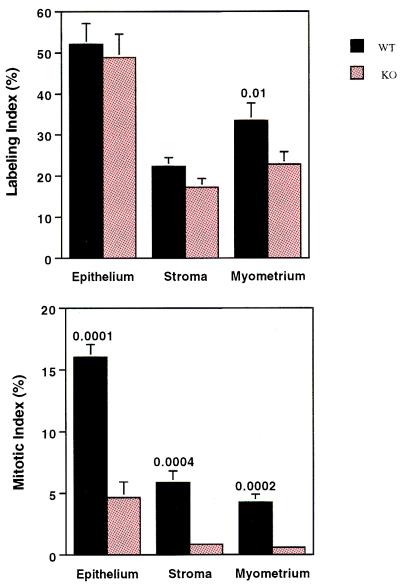
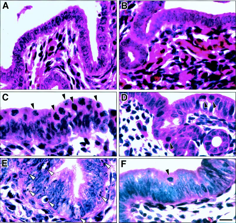
Insulin-like growth factor 1 (IGF1) has been proposed as a "G1-progression factor" and as a mediator of estradiol's (E2) mitogenic effects on the uterus. To test these hypotheses, we compared E2's mitogenic effects on the uteri of Igf1-targeted gene deletion (null) and wild-type littermate mice. The proportion of uterine cells involved in the cell cycle and G1- and S-phase kinetics were not significantly different in wild-type and Igf1-null mice. However, the appearance of E2-induced mitotic figures and cell number increases were profoundly retarded in Igf1-null uterine tissue. There was a significant increase in nuclear DNA concentration in Igf1-null cells, consistent with a G2 arrest. Interestingly, apoptotic cells were also significantly reduced in abundance, and the normal massive apoptotic response to E2 withdrawal was absent in the Igf1-null uterus. These data show that Igf1 is an essential mediator of E2's mitogenic effects, with a critical role not in G1 progression but in G2 progression.


Figures

Figure 1
Figure 1
Autoradiographic visualization of E2-induced DNA synthesis in wt (A, C, and E) and Igf1-null (B, D, and F) uteri. A and C and B and D are paired bright- and dark-field low-magnification photomicrographs. E and F are higher magnification dark-field views showing localization of tritium-positive red nuclei in epithelium (ep), stroma (st), and myometrium (my). Mice were given a single intraperitoneal injection of 17-β-estradiol (1 μg/g) at 0 h and injected with [3H]thymidine (2 μCi/g; 1 Ci = 37 GBq) 1 h before sacrifice at 21 h. Sections from the middle third of each uterine horn were exposed to photographic emulsion for 3 weeks, developed, and stained with hematoxylin/eosin and photographed through a red filter. Magnification ×50 (AD) and ×200 (E and F).
Figure 2
Figure 2
To compare S-phase duration and flux in wt and Igf1−/− uteri, mice were given [3H]thymidine (2 μCi/g) 3 h before and BrdUrd (Amersham Pharmacia RPN 201, 10 μl/g) 1 h before sacrifice. Uterine sections were processed for immunodetection of BrdUrd and then for autoradiographic detection of 3H-labeled nuclei. 3H-only-positive nuclei (>5 silver grains) are indicated by solid arrowheads, BrdUrd-only-positive nuclei by open arrowheads, and doubly labeled nuclei by twin arrowheads. wt (A) and Igf1-null (B) uteri demonstrate equal proportions of cells in each category (see Table 2). 3H-only-positive nuclei represent cells that exited S phase and BrdUrd-only positive nuclei represent cells that entered S phase during the course of the experiment. Doubly labeled nuclei represent cells that were in S phase for over 2 h (the time between thymidine and BrdUrd injection) during the course of the experiment. The percentage of doubly labeled cells reflects of S-phase duration. (Bar = 25 μm.)
Figure 3
Figure 3
Comparison of labeling index and mitotic index in the different cell types of wt and Igf1-null uteri 21 h after E2 stimulation. These data are from P20 mice, at which age the greatest proliferative response to E2 occurs. Similar data (14.7 ± 2.1 vs. 4.0 ± 1.3, respectively) were obtained from the analysis of the mitotic index in P40 wt and Igf1-null uterine epithelium. The mitotic index in other uterine cell types at P40 was so low in both wt and Igf1-null uteri that it was not counted. The values shown are means ± SEM for 8–12 mice per group.
Figure 4
Figure 4
Mitosis and apoptosis in wt (A, C, and E) and Igf1-null (B, D, and F) uterine epithelium. Mice were treated with vehicle (A and B) or E2 for 21 h (C and D) or 48 h (E and F). Solid arrowheads indicate some mitotic figures, and open arrowheads show examples of apoptotic bodies. The significantly greater occurrence of apoptosis in wt uteri shown here by hematoxylin/eosin staining was confirmed by analysis of DAPI-stained sections and by in situ end-labeling of fragmented DNA. (Bar = 10 μm.)

References

    1. Murphy L J, Ghahary A. Endocr Rev. 1990;11:443–455. - PubMed
    1. Sirbasku D A. Proc Natl Acad Sci USA. 1978;75:3786–3789. - PMC - PubMed
    1. Murphy L J, Murphy L C, Freisen H G. Endocrinology. 1987;1:445–451. - PubMed
    1. Norstedt G, Levinovitz A, Erikson H. Acta Endocrinol. 1989;120:466–473. - PubMed
    1. Kapur S, Tamada H, Dey S K, Andrews G K. Biol Reprod. 1992;46:208–211. - PubMed

Publication types

LinkOut - more resources