. 1999 Mar;103(6):779-88.
doi: 10.1172/JCI5909.

Pulmonary expression of interleukin-13 causes inflammation, mucus hypersecretion, subepithelial fibrosis, physiologic abnormalities, and eotaxin production

Affiliations

Pulmonary expression of interleukin-13 causes inflammation, mucus hypersecretion, subepithelial fibrosis, physiologic abnormalities, and eotaxin production

Z Zhu et al. J Clin Invest. 1999 Mar.

Abstract

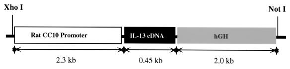
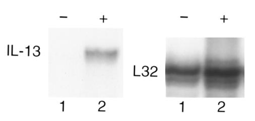
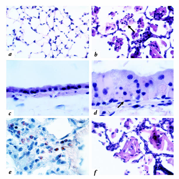
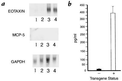
Interleukin (IL)-13 is a pleiotropic cytokine produced in large quantities by activated CD4(+) Th2 lymphocytes. To define further its potential in vivo effector functions, the Clara cell 10-kDa protein promoter was used to express IL-13 selectively in the lung, and the phenotype of the resulting transgenic mice was characterized. In contrast to transgene-negative littermates, the lungs of transgene-positive mice contained an inflammatory response around small and large airways and in the surrounding parenchyma. It was mononuclear in nature and contained significant numbers of eosinophils and enlarged and occasionally multinucleated macrophages. Airway epithelial cell hypertrophy, mucus cell metaplasia, the hyperproduction of neutral and acidic mucus, the deposition of Charcot-Leyden-like crystals, and subepithelial airway fibrosis were also prominently noted. Eotaxin protein and mRNA were also present in large quantities in the lungs of the transgene-positive, but not the transgene-negative, mice. IL-4, IL-5, granulocyte-macrophage colony-stimulating factor, and monocyte chemoattractant protein-5 were not similarly detected. Physiological evaluations revealed significant increases in baseline airways resistance and airways hyperresponsiveness (AHR) to methacholine in transgene-positive animals. Thus, the targeted pulmonary expression of IL-13 causes a mononuclear and eosinophilic inflammatory response, mucus cell metaplasia, the deposition of Charcot-Leyden-like crystals, airway fibrosis, eotaxin production, airways obstruction, and nonspecific AHR. IL-13 may play an important role in the pathogenesis of similar responses in asthma or other Th2-polarized tissue responses.


Figures

Figure 1
Figure 1
Schematic illustration of the construct in the IL-13 transgenic mice. hGH, human growth hormone; IL, interleukin.
Figure 2
Figure 2
Ribonuclease protection analysis of IL-13 gene expression in transgene (+) mice. Total RNA was isolated from the lungs from transgene (+) (lane 2) and () mice, and the levels of IL-13 and L32 mRNA were quantitated by ribonuclease protection as described in Methods.
Figure 3
Figure 3
Comparison of the histological features of transgene (+) and (–) mice. Lungs from littermate transgene (–) and (+) mice were obtained, fixed, sectioned, and stained. (a) Transgene (–) lung parenchyma. H&E, ×100 (all photographs are original magnification). (b) Transgene (+) lung parenchyma. H&E, ×100. (c) Transgene (–) airway wall and epithelium. H&E, ×250. (d) Transgene (+) airway wall and epithelium. H&E, ×250. (e) Transgene (+) lung parenchyma. Congo red, ×250. (f) Transgene (+) lung parenchyma. H&E, ×100. A comparison of a and b illustrates the inflammatory response, macrophage enlargement, and collections of eosinophils (b, arrow) in the transgene (+) animals. A comparison of c and d illustrates the epithelial cell hypertrophy and intracellular refractile material (d, arrow) in the transgene (+) animals. e illustrates the tissue eosinophils (small red cells with bilobed nuclei). f illustrates the alveolar crystals in the transgene (+) animals. Eosinophils and crystals were not seen in the lungs from the transgene (–) animals. H&E, hematoxylin and eosin.
Figure 4
Figure 4
Comparison of mucus production by airway epithelium in transgene (–) and (+) animals. Lungs from littermate transgene (–) (a and c) and (+) (b and d) animals were obtained, fixed, and subjected to PAS (a and b) and alcian blue (c and d) staining. ×100. PAS and alcian blue staining cells have purple and blue cytoplasmic inclusions, respectively. PAS, periodic acid-Schiff.
Figure 5
Figure 5
Comparison of the airway collagen in transgene (–) and (+) animals. Lungs from transgene (–) (a and c) and (+) (b and d) animals were obtained, fixed, sectioned, and stained (Masson's trichrome). The blue staining collagen in the small airways (a and b; ×100) and large airways (c and d; ×250) are compared.
Figure 6
Figure 6
Documentation of eotaxin production in lungs from transgene (+) mice. (a) Illustrates the levels of eotaxin, monocyte chemoattractant protein-5 (MCP-5), and glyceraldehyde phosphate dehydrogenase (GAPDH) mRNA in total lung RNA from transgene (–) (lanes 1 and 2) and (+) (lanes 3 and 4) animals (assessed using Northern analysis). Each lane contains RNA from a separate animal. (b) Illustrates the levels of eotaxin protein detected by ELISA in BAL fluid from transgene (–) and (+) mice. Each value represents the mean ± SE of assays involving four mice (P < 0.001). BAL, bronchoalveolar lavage.
Figure 7
Figure 7
Comparison of the airway physiology of 1-month-old transgene (–) and line 2 transgene (+) mice. The airways resistance of transgene (–) (open triangles) and (+) (filled triangles) littermate mice was evaluated before and at intervals after methacholine administration using noninvasive methodology as described in the Methods. Enhanced pause (Penh) is expressed as percent increase over baseline values. The noted values represent the mean ± SEM of assays on a minimum of five animals. *P < 0.01.

References

    1. Lucey DR, Clerici M, Shearer GM. Type 1 and type 2 cytokine dysregulation in human infectious, neoplastic, and inflammatory diseases [review] Clin Microbiol Rev. 1996;9:532–562. - PMC - PubMed
    1. Abbas AK, Murphy KM, Sher A. Functional diversity of helper T lymphocytes. Nature. 1996;383:787–793. - PubMed
    1. Mosmann TR, Sad S. The expanding universe of T-cell subsets: Th1, Th2 and more. Immunol Today. 1996;17:138–146. - PubMed
    1. Moller DR, et al. Enhanced expression of IL-12 associated with Th1 cytokine profiles in active pulmonary sarcoidosis. J Immunol. 1996;156:4952–4960. - PubMed
    1. Barner M, Mohrs M, Brombacher F, Kopf M. Differences between IL-4R alpha–deficient and IL-4–deficient mice reveal a role for IL-13 in the regulation of Th2 responses. Curr Biol. 1998;8:669–672. - PubMed

MeSH terms