. 1999 Mar;103(6):851-8.
doi: 10.1172/JCI5257.

Selective activation and functional significance of p38alpha mitogen-activated protein kinase in lipopolysaccharide-stimulated neutrophils

Affiliations

Selective activation and functional significance of p38alpha mitogen-activated protein kinase in lipopolysaccharide-stimulated neutrophils

J A Nick et al. J Clin Invest. 1999 Mar.

Abstract

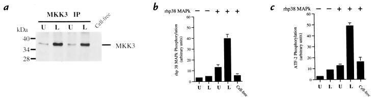
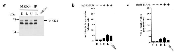
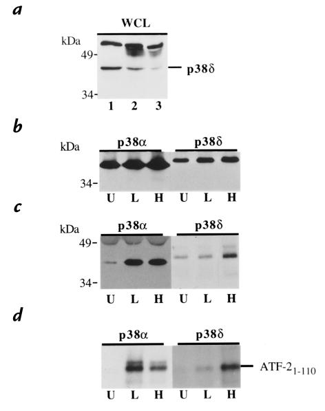
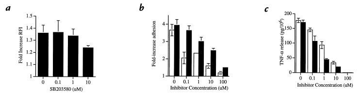
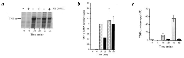
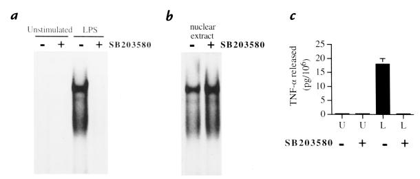
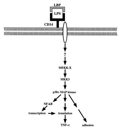
Activation of leukocytes by proinflammatory stimuli selectively initiates intracellular signal transduction via sequential phosphorylation of kinases. Lipopolysaccharide (LPS) stimulation of human neutrophils is known to result in activation of p38 mitogen-activated protein kinase (MAPk); however, the upstream activator(s) of p38 MAPk is unknown, and consequences of p38 MAPk activation remain largely undefined. We investigated the MAPk kinase (MKK) that activates p38 MAPk in response to LPS, the p38 MAPk isoforms that are activated as part of this pathway, and the functional responses affected by p38 MAPk activation. Although MKK3, MKK4, and MKK6 all activated p38 MAPk in experimental models, only MKK3 was found to activate recombinant p38 MAPk in LPS-treated neutrophils. Of p38 MAPk isoforms studied, only p38alpha and p38delta were detected in neutrophils. LPS stimulation selectively activated p38alpha. Specific inhibitors of p38alpha MAPk blocked LPS-induced adhesion, nuclear factor-kappa B (NF-kappaB) activation, and synthesis of tumor necrosis factor-alpha (TNF-alpha). Inhibition of p38alpha MAPk resulted in a transient decrease in TNF-alpha mRNA accumulation but persistent loss of TNF-alpha synthesis. These findings support a pathway by which LPS stimulation of neutrophils results in activation of MKK3, which in turn activates p38alpha MAPk, ultimately regulating adhesion, NF-kappaB activation, enhanced gene expression of TNF-alpha, and regulation of TNF-alpha synthesis.


Figures

Figure 1
Figure 1
Coupled assay of MKK3 activation. Neutrophils were stimulated with LPS (100 ng/ml) for 20 min at 37°C. (a) Phosphorylation of MKK3. MKK3 was immunoprecipitated from cell lysates of LPS-stimulated (L) or unstimulated (U) neutrophils. Lysates were submitted to SDS-PAGE and Western blotting with an anti-phosphorylated MKK3 antibody. A cell-free mixture that did not contain immunoprecipitated MKK3 is shown to control for intrinsic activity of the rhp38 MAPk. Blot is representative of three experiments. (b) Activation of MKK3. 32P phosphorylation of rhp38 MAPk from blots was quantified by phosphor screen autoradiography. Lysates in the absence of rhp38 MAPk (lanes 1 and 2) were compared with lysates in the presence of rhp38 MAPk (lanes 3 and 4) and a cell-free control to demonstrate specific activation of MKK3 in response to LPS as determined by 32P phosphorylation of rhp38 MAPk. (c) Coupled activation of rhp38 MAPk by activated MKK3. The ability of activated MKK3 to activate rhp38 MAPk was determined by the ability of phosphorylated rhp38 MAPk to phosphorylate the substrate ATF-21-110. 32P phosphorylation of ATF-21-110 from blots was quantified by phosphor screen autoradiography. Lysates in the absence of rhp38 MAPk (lanes 1 and 2) represent baseline phosphorylation of the ATF-21-110, whereas lysates in the presence of rhp38 MAPk (lanes 3 and 4) demonstrate increased phosphorylation of ATF-21-110 via coupled activation of rhp38 MAPk by activated MKK3 in response to LPS. The cell-free control quantifies intrinsic activity of the rhp38 MAPk and is equivalent to the unstimulated lysate in lane 3. Plots depict mean values and SEM from three consecutive experiments expressed in arbitrary units. ATF, activated transcription factor; LPS, lipopolysaccharide; MAPk, mitogen-activated protein kinase; MKK, MAPk kinase.
Figure 2
Figure 2
Coupled assay of MKK4 activation. Neutrophils were stimulated with LPS (100 ng/ml) for 20 min at 37°C. (a) Phosphorylation of MKK4. Under identical conditions to the experiments shown in Fig. 1, MKK4 was immunoprecipitated from cell lysates and submitted to SDS-PAGE and Western blotting with an anti-phosphorylated MKK4 antibody. Blot is representative of three experiments. (b) Activation of MKK4. 32P phosphorylation of rhp38 MAPk from blots was quantified by phosphor screen autoradiography. Lysates in the absence of rhp38 MAPk (lanes 1 and 2) were compared with lysates in the presence of rhp38 MAPk (lanes 3 and 4) and a cell-free control to demonstrate the lack of specific activation of MKK4 in response to LPS as determined by 32P phosphorylation of rhp38 MAPk. (c) Coupled activation of rhp38 MAPk by MKK4. The ability of MKK4 to activate rhp38 MAPk was determined by the ability of phosphorylated rhp38 MAPk to phosphorylate the substrate ATF-21-110. Lysates in the absence of rhp38 MAPk (lanes 1 and 2) represent baseline phosphorylation of the ATF-21-110, whereas lysates in the presence of rhp38 MAPk (lanes 3 and 4) demonstrate little increase in phosphorylation of ATF-21-110 via coupled activation of rhp38 MAPk by MKK4 in response to LPS. The cell-free control quantifies intrinsic activity of the rhp38 MAPk and is equivalent to the unstimulated and LPS-stimulated lysates in lanes 3 and 4. Plots depict mean values and SEM from three consecutive experiments expressed in arbitrary units.
Figure 3
Figure 3
LPS stimulation selectively activates p38α MAPk, but not p38δ MAPk. (a) Identification of p38δ MAPk in neutrophil whole-cell lysates (WCL) by immunodepletion. Neutrophil lysate were subjected to SDS-PAGE and Western blotting with anti–peptide p38δ antisera (lane 1). Confirmation of the identity of the p38δ MAPk band was achieved by immunoprecipitating p38δ from the lysate using a purified anti–full-length p38δ antibody. A significant decrease in the proposed p38δ band is observed after 6 h (lane 2) and 12 h (lane 3) of immunodepletion. (b) Immunoprecipitation of p38α and p38δ MAPk. Neutrophils stimulated with LPS (100 ng/ml) for 25 min at 37°C (L), H2O2 (1 mM) for 20 min at 37°C (H), or unstimulated (U) were lysed, and p38α and p38δ MAPk were immunoprecipitated and separated by SDS-PAGE. Western blots were probed with antisera specific for p38α and p38δ MAPk to demonstrate equivalent amounts of immunoprecipitation for each condition studied. (c) Tyrosine phosphorylation of p38α and p38δ MAPk. Blots from b were reprobed with an anti-phosphotyrosine antibody capable of reacting with phosphorylated tyrosine residues from both p38α and p38δ MAPk. (d) Activation of p38α and p38δ MAPk. Neutrophils subjected to identical conditions as in b and c were lysed, and p38α and p38δ MAPk was immunoprecipitated and combined with ATF-21-110 in the presence of [32P]ATP. The peptide was subjected to SDS-PAGE, and the degree of 32P phosphorylation of ATF-21-110 was assessed by autoradiography of the blot. Each panel is representative of three consecutive experiments.
Figure 4
Figure 4
Effect of p38α MAPk inhibition on LPS-induced rapid neutrophil responses. Neutrophils suspended in SK&F86002 (open bars) or SB203580 (filled bars) over a range of concentrations for 60 min at 37°C were then stimulated with LPS (100 ng/ml) at 37°C. (a) Effect of in vivo inhibition of p38α MAPk on actin assembly. The -fold increase in relative fluorescence index (RFI) of LPS-stimulated neutrophils (5 min) compared with unstimulated cells was plotted for each concentration of SB203580. The plot represents mean activity and SEM for three independent experiments. The relationship between SB203580 concentration and inhibition of actin assembly was not statistically significant (P = 0.55). (b) Effect of in vivo inhibition of p38α MAPk on neutrophil adhesion. The -fold increase in adhesion of LPS-stimulated neutrophils (30 min) compared with unstimulated cells was plotted for each concentration of inhibitor. The plot represents mean values and SEM for six experiments. The relationship between the concentrations of SK&F86002 or SB203580 and inhibition of adhesion is significant (P < 0.0001). (c) Effect of p38α MAPk inhibition on LPS-induced release of TNF-α. The quantity of TNF-α released per 106 neutrophils stimulated with LPS (120 min) was plotted for each concentration of inhibitor. The plot represents mean values and SEM of three consecutive experiments. The relationship between the concentrations of SK&F86002 or SB203580 and inhibition of TNF-α release is significant (P < 0.0001). TNF, tumor necrosis factor.
Figure 5
Figure 5
Quantification of TNF-α mRNA by RPA. (a) RPA autoradiograph of TNF-α mRNA at 0, 30, and 60 min after stimulation with LPS (100 ng/ml) at 37°C in the presence (+) and absence () of SB203580 (10 μM). An increase in TNF-α mRNA seen after 30 min of stimulation (lane 3) is substantially reduced in cells treated with SB203580 (lane 4). At 60 min, the quantity of TNF-α mRNA in the untreated and SB203580-treated cells (lanes 5 and 6) is equivalent. The blot is representative of three consecutive experiments. (b) Normalized plot of TNF-α mRNA synthesis as quantified by RPA. Amount of TNF-α mRNA present for each condition was expressed as a fraction of GAPDH present for each sample to correct for potential differences in sample loading. Each value was then normalize to the amount of TNF-α mRNA present at 30 min of LPS stimulation in the untreated neutrophils (lane 3) to correct for variability in response between donors. After 30 min of LPS stimulation, SB203580-treated neutrophils (closed bar) demonstrated 42% of the TNF-α mRNA present in the untreated cells (hatched bar). After 60 min of stimulation, no significant difference is seen. The panel represents mean values and SEM of three consecutive experiments. (c) Release of TNF-α under conditions studied. TNF-α released from the neutrophils studied in b was assayed. The quantity of TNF-α released per 106 neutrophils was plotted for each time in the presence (closed bars) or absence (hatched bars) of SB203580. The panel represents mean values and SEM of the three consecutive experiments described for b. GAPDH, glyceraldehyde phosphate dehydrogenase; RPA, RNase protection assay.
Figure 6
Figure 6
Inhibition of NF-κB activation by SB203580-induced inhibition of p38α MAPk. (a) Effect of in vivo inhibition of p38α MAPk on NF-κB activation. Neutrophils were suspended in the presence or absence of SB203580 (10 μM) for 60 min at 37°C and subsequently stimulated with LPS (100 ng/ml) for 20 min at 37°C or left unstimulated. Nuclear extracts were then prepared and analyzed in EMSA using an NF-κB oligonucleotide probe. This blot is representative of three consecutive experiments. (b) Lack of a direct effect of SB203580 toward the binding of NF-κB to its cognate sequence. Nuclear extracts from LPS-stimulated neutrophils were coincubated with SB203580 (100 μM) for 30 min at room temperature in the binding mixture, before EMSA analysis. This blot is representative of three consecutive experiments. (c) Release of TNF-α at the time of NF-κB activation. The quantity of TNF-α released per 106 neutrophils from the samples studied in a was plotted for each condition. Bars depict the mean value and SEM of TNF-α release (per 106 neutrophils) from three independent experiments.EMSA, electrophoretic mobility shift assay; NF-κB, nuclear factor-kappa B.
Figure 7
Figure 7
Scheme depicting proposed intracellular signaling pathways and functional consequences in response to stimulation of human neutrophils with LPS. Binding of CD14 by LPS in the presence of LBP initiates a signal that passes though a transmembrane spanning protein and a series of yet unknown upstream signaling events, leading to activation of the MKK3–p38α MAPk cascade. The activator of MKK3 in this cell (MEKK-X) is not known. Activation of p38α MAPk then results in rapid responses such as adhesion and activation of NF-κB. Synthesis of the TNF-α peptide is also dependent on p38α MAPk activation, in part through activation of NF-κB, but primarily through regulation of translation. LBS, lipopolysaccharide-binding protein.

Similar articles

Cited by

References

    1. Cobb MH, Goldsmith EJ. How MAP kinases are regulated. J Biol Chem. 1995;270:14843–14846. - PubMed
    1. Han J, Lee J-D, Bibbs L, Ulevitch RJ. A MAP kinase targeted by endotoxin and hyperosmolarity in mammalian cells. Science. 1994;265:808–811. - PubMed
    1. Rouse J, et al. A novel kinase cascade triggered by chemical stress and heat shock which stimulates MAPKAP-2 and phosphorylation of the small heat shock proteins. Cell. 1994;78:1027–1037. - PubMed
    1. Lee JC, et al. A protein kinase involved in the regulation of inflammatory cytokine biosynthesis. Nature. 1994;372:739–746. - PubMed
    1. Nick JA, et al. Activation of a p38 MAPk in human neutrophils by LPS. J Immunol. 1996;156:4867–4875. - PubMed

Publication types

MeSH terms

Substances