. 1999 Mar;103(6):879-87.
doi: 10.1172/JCI6042.

Protease-activated receptors 1 and 4 mediate activation of human platelets by thrombin

Affiliations

Protease-activated receptors 1 and 4 mediate activation of human platelets by thrombin

M L Kahn et al. J Clin Invest. 1999 Mar.

Abstract

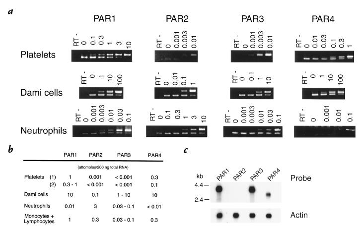
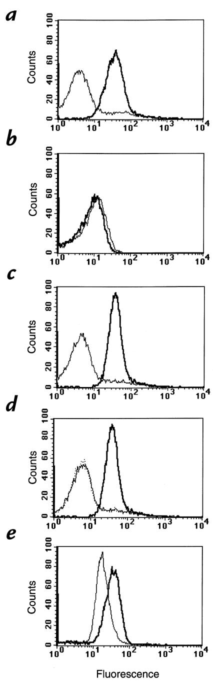
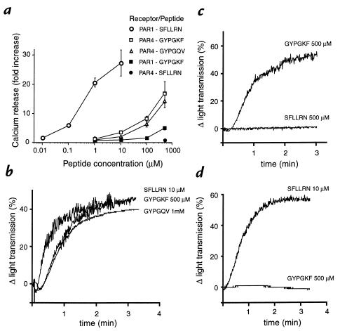
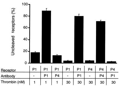
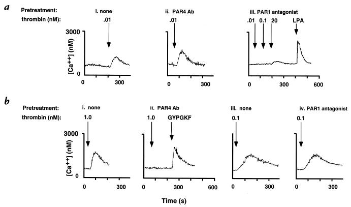
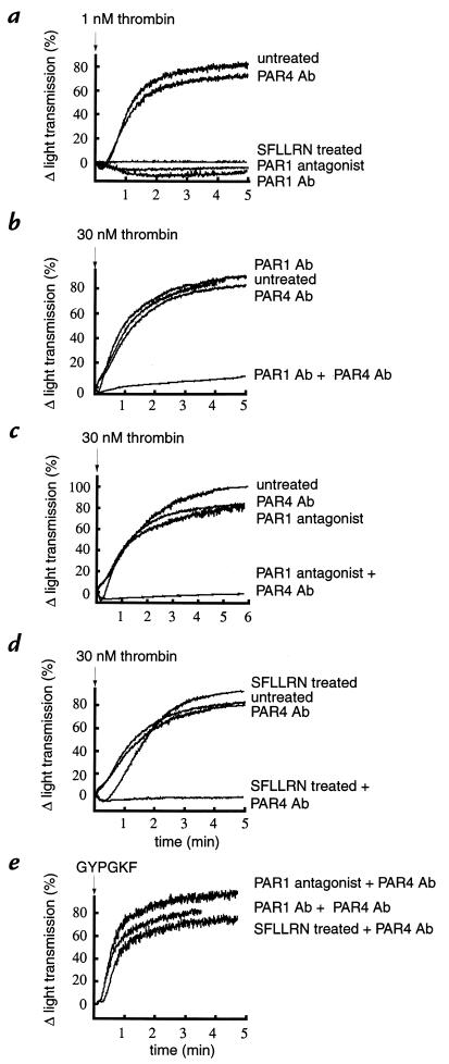
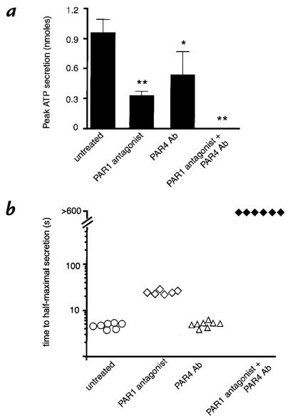
Because of the role of thrombin and platelets in myocardial infarction and other pathological processes, identifying and blocking the receptors by which thrombin activates platelets has been an important goal. Three protease-activated receptors (PARs) for thrombin -- PAR1, PAR3, and PAR4 -- are now known. PAR1 functions in human platelets, and the recent observation that a PAR4-activating peptide activates human platelets suggests that PAR4 also acts in these cells. Whether PAR1 and PAR4 account for activation of human platelets by thrombin, or whether PAR3 or still other receptors contribute, is unknown. We have examined the roles of PAR1, PAR3, and PAR4 in platelets. PAR1 and PAR4 mRNA and protein were detected in human platelets. Activation of either receptor was sufficient to trigger platelet secretion and aggregation. Inhibition of PAR1 alone by antagonist, blocking antibody, or desensitization blocked platelet activation by 1 nM thrombin but only modestly attenuated platelet activation by 30 nM thrombin. Inhibition of PAR4 alone using a blocking antibody had little effect at either thrombin concentration. Strikingly, simultaneous inhibition of both PAR1 and PAR4 virtually ablated platelet secretion and aggregation, even at 30 nM thrombin. These observations suggest that PAR1 and PAR4 account for most, if not all, thrombin signaling in platelets and that antagonists that block these receptors might be useful antithrombotic agents.


Figures

Figure 1
Figure 1
Expression of mRNAs encoding PAR1, PAR2, PAR3, and PAR4 in platelets, Dami cells, and neutrophils. (a) Competitive RT-PCR of total RNA from platelets, Dami cells, and neutrophils. Total cellular RNA (200 ng) mixed with the indicated quantity of competitor RNA (measured in attomoles) was reverse-transcribed and amplified. Products were digested with a restriction endonuclease to distinguish the products of competitor RNA (uncleavable upper band) vs. native cellular mRNA (lower band). RT indicates mock RT-PCR of total cellular RNA and the highest amount of competitor RNA with no reverse transcriptase added. Each sample was analyzed at least twice. Note that the single band seen in the platelet PAR3 RT-PCR is due to amplification of competitor RNA. (b) Quantitation of PAR mRNAs in platelet, Dami cell, neutrophil, and monocyte/lymphocyte preparations. Results indicate the range of values obtained from at least two experiments like that shown in a. 1 amol/200 ng corresponds to an mRNA relative abundance of roughly 1:3,000. The expression of PAR mRNA in the platelets of two unrelated individuals is shown. (c) Northern blot analysis of PAR gene expression in Dami cells. Blots were hybridized separately with coding region probes for PAR1, PAR2, PAR3, or PAR4, as well as with probe for β-actin mRNA as a control for lane loading. Note concordance with PCR data in b. PAR, protease-activated receptor; RT, reverse transcription.
Figure 2
Figure 2
Flow cytometric analysis of platelets for surface expression of PAR1, PAR3, and PAR4. Fixed platelets were incubated with preimmune IgG (narrow lines) or PAR1 IgG (a), PAR3 IgG (b), or PAR4 IgG (c) (wide lines) and then analyzed as described in Methods. (d) Platelets were incubated with PAR4 IgG in the absence (wide line) or presence (thin line) of the peptide antigen (1 μM) used to generate the PAR4 antiserum, or after treatment with 20 nM thrombin for 10 min at 37°C (dotted line). Each curve represents an analysis of 10,000 events. This experiment was repeated twice with separate donors with equivalent results. (e) Flow cytometric analysis of Dami cells as a positive control for detection of PAR3. Fixed Dami cells were incubated with preimmune IgG (narrow line) or PAR3 IgG (wide line) and then analyzed as above. Dami cells were also positive for PAR1 and PAR4 (not shown).
Figure 3
Figure 3
Effects of PAR1- and PAR4-activating peptides. (a) Specificity and potency. Peptide-triggered 45Ca release was measured in Xenopus oocytes expressing human PAR1 and human PAR4 tagged at their NH2-termini with a FLAG epitope. Data are mean ± SEM (n = 3) and are expressed as fold increase over baseline for each receptor. Surface expression of PAR1 measured with anti-FLAG monoclonal antibody was 1.3 times that of PAR4. This experiment was replicated twice. (bd) Activation of human platelets with the PAR1-activating peptide SFLLRN and the PAR4-activating peptides GYPGKF and GYPGQV. (b) Platelets were exposed to either SFLLRN (10 μM) or GYPGKF (500 μM) or GYPGQV (1 mM) at time 0, and aggregation was measured as change in light transmission. (c) SFLLRN-desensitized platelets (see Methods) were exposed to either SFLLRN (500 μM) or GYPGKF (500 μM) at time 0, and aggregation was measured as change in light transmission. (d) GYPGKF-desensitized platelets (see Methods) were exposed to either SFLLRN (500 μM) or GYPGKF (500 μM) at time 0, and aggregation was measured as change in light transmission. The experiments in b, c, and d were repeated three times.
Figure 4
Figure 4
Inhibition of thrombin cleavage of receptor NH2-terminus by anti-PAR1 and anti-PAR4 antibodies. Rat-1 cells expressing PAR1 and PAR4 bearing the FLAG epitope at their extreme NH2-termini were fixed and then incubated with PAR1 IgG (P1; 100 μg/ml), PAR4 IgG (P4; 1 mg/ml), or buffer alone for 60 min before exposure to either 1 or 30 nM thrombin for 10 min at 37°C. Receptor cleavage was measured as loss of binding sites for M1 monoclonal antibody to the FLAG epitope, which was NH2-terminal to the thrombin cleavage site in both receptors, so as to be lost from the cells upon receptor cleavage. Data (mean ± SEM; n = 3) are expressed as percent of control cells exposed to buffer alone. This experiment was repeated twice.
Figure 5
Figure 5
Inhibition of PAR1 and PAR4 signaling by PAR1- and PAR4-blocking antibodies and PAR1 antagonist. Fibroblast cell lines in which thrombin signaling was mediated solely by PAR1 (a) or by PAR4 (b) were incubated with buffer alone (none), PAR4 IgG (PAR4 Ab; 1 mg/ml), or the PAR1 antagonist BMS200261 (100 μM) for 30 min at 37°C. Cells were then exposed to thrombin (0.01, 0.1, 1.0, or 20 nM as indicated), GYPGKF (500 μM), or lysophosphatidic acid (LPA; 5 μM). Receptor-triggered increases in cytoplasmic calcium were measured fluorometrically using the calcium sensitive dye Fura-2. This experiment was repeated three times with similar results. Ab, antibody.
Figure 6
Figure 6
The effects of inhibition of PAR1 and/or PAR4 on aggregation of human platelets in response to low (1 nM) and high (30 nM) concentrations of thrombin. Platelets were pretreated with buffer alone, PAR1 IgG (10 μg/ml), PAR4 IgG (1 mg/ml), or PAR1 antagonist (100 μM), or were desensitized to SFLLRN as indicated and then exposed to 1 nM thrombin (a), 30 nM thrombin (bd), or 500 μM GYPGKF (e) at time 0. Aggregation was measured as increase in light transmission. Preimmune or nonimmune IgG were without effect (not shown). This experiment was performed using triplicate samples twice (a, c, e) or four times (b, d). Representative tracings are shown.
Figure 7
Figure 7
The effects of inhibition of PAR1 and/or PAR4 on platelet ATP secretion in response to thrombin. (a) Peak ATP secretion. Platelets were pretreated with buffer alone, PAR4 IgG (1 mg/ml), PAR1 antagonist BMS200261 (100 μM), or PAR1 antagonist plus PAR4 IgG as indicated, and then stimulated with 30 nM thrombin. Peak ATP concentration in the 10 min after addition of thrombin was measured by lumiaggregometry. Preimmune IgG had no effect (not shown). Data are mean ± SD (n = 5–7) of peak secretion measured; similar results were obtained with platelets from two individuals. Data were analyzed by two-way ANOVA and t test with a Bonferroni correction for multiple comparisons. *P ≅ 0.06, **P < 0.001 compared with untreated group. Note that no secretion was detected during the 10 min after addition of 30 nM thrombin to platelets treated with PAR1 antagonist plus PAR4 IgG. (b) Time to half-maximal secretion. Time to reach 50% of the peak ATP secretion response elicited by 30 nM thrombin in each group (a) was measured. Platelets were pretreated with buffer alone (open circles), PAR1 antagonist (open diamonds), PAR4 IgG (open triangles), or PAR1 antagonist plus PAR4 Ab (closed diamonds) as in a, and then stimulated with 30 nM thrombin. Points displayed as >600 s indicate no measurable secretion within 10 min after addition of thrombin. PAR4 preimmune IgG had no effect inhibitory effect in such experiments, even in the presence of PAR1 antagonist (not shown).

References

    1. Davey M, Luscher E. Actions of thrombin and other coagulant and proteolytic enzymes on blood platelets. Nature. 1967;216:857–858. - PubMed
    1. Berndt, M., and Phillips, D. 1981. Platelet membrane proteins: composition and receptor function. In Platelets in biology and pathology. J.L. Gordon, editor. Elsevier. Amsterdam, the Netherlands. 43–74.
    1. Vu T-KH, Hung DT, Wheaton VI, Coughlin SR. Molecular cloning of a functional thrombin receptor reveals a novel proteolytic mechanism of receptor activation. Cell. 1991;64:1057–1068. - PubMed
    1. Rasmussen UB, et al. cDNA cloning and expression of a hamster alpha-thrombin receptor coupled to Ca2+ mobilization. FEBS Lett. 1991;288:123–128. - PubMed
    1. Vu T-KH, Wheaton VI, Hung DT, Coughlin SR. Domains specifying thrombin-receptor interaction. Nature. 1991;353:674–677. - PubMed

Publication types