. 1999 Mar;103(6):907-14.
doi: 10.1172/JCI5578.

Targeted mutation of plasma phospholipid transfer protein gene markedly reduces high-density lipoprotein levels

Affiliations

Targeted mutation of plasma phospholipid transfer protein gene markedly reduces high-density lipoprotein levels

X C Jiang et al. J Clin Invest. 1999 Mar.

Abstract

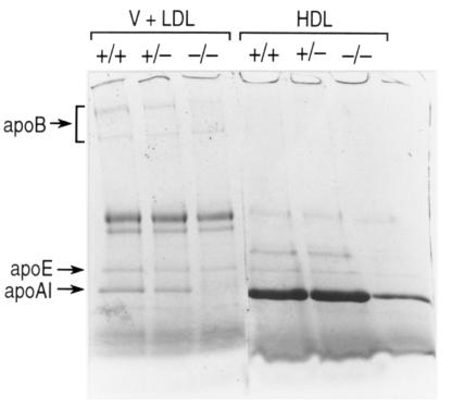
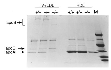
It has been proposed that the plasma phospholipid transfer protein (PLTP) facilitates the transfer of phospholipids and cholesterol from triglyceride-rich lipoproteins (TRL) into high-density lipoproteins (HDL). To evaluate the in vivo role of PLTP in lipoprotein metabolism, we used homologous recombination in embryonic stem cells and produced mice with no PLTP gene expression. Analysis of plasma of F2 homozygous PLTP-/- mice showed complete loss of phosphatidylcholine, phosphatidylethanolamine, phosphatidylinositol, sphingomyelin, and partial loss of free cholesterol transfer activities. Moreover, the in vivo transfer of [3H]phosphatidylcholine ether from very-low-density proteins (VLDL) to HDL was abolished in PLTP-/- mice. On a chow diet, PLTP-/- mice showed marked decreases in HDL phospholipid (60%), cholesterol (65%), and apo AI (85%), but no significant change in non-HDL lipid or apo B levels, compared with wild-type littermates. On a high-fat diet, HDL levels were similarly decreased, but there was also an increase in VLDL and LDL phospholipids (210%), free cholesterol (60%), and cholesteryl ester (40%) without change in apo B levels, suggesting accumulation of surface components of TRL. Vesicular lipoproteins were shown by negative-stain electron microscopy of the free cholesterol- and phospholipid-enriched IDL/LDL fraction. Thus, PLTP is the major factor facilitating transfer of VLDL phospholipid into HDL. Reduced plasma PLTP activity causes markedly decreased HDL lipid and apoprotein, demonstrating the importance of transfer of surface components of TRL in the maintenance of HDL levels. Vesicular lipoproteins accumulating in PLTP-/- mice on a high-fat diet could influence the development of atherosclerosis.


Figures

Figure 1
Figure 1
Strategy used to disrupt the mouse PLTP gene. (a) The top line represents the map of the endogenous murine PLTP gene and its flanking sequence. The middle line represents the vector used to target the PLTP locus. The bottom line shows the predicted organization of the locus after homologous recombination. A probe and a pair of PCR primers indicated in this line were used to confirm the integrity of site-specific integration. (b) Southern blot analysis of ES cells and (c) mouse tail-tip genomic DNA, respectively, digested with EcoRI and hybridized with the probe. Normal ES cell DNA and control mice DNA with 2.1-kb signal only (+/+); targeted ES cell DNA and heterozygous deficient mice with 2.1-kb and 1.8-kb signal (+/–); homozygous deficient mice with 1.8-kb signal only (–/–). ES, embryonic stem; neo, neomycin-resistant gene; PLTP, phospholipid transfer protein; tK, herpes simplex virus thymidine kinase gene.
Figure 2
Figure 2
PLTP mRNA measurement. (a) Northern blot of poly(A)+ RNA (2 μg) from lung probed with a mouse PLTP cDNA (666 bp, nucleotides 3–669). (b) RNase protection analysis of mouse tissues. A fragment of the mouse PLTP cDNA (160 bp, codons 750–910) was cloned into Bluescript KS+ (Stratagene, La Jolla, California, USA) and used to prepare radiolabeled cRNA probes; 30 μg of total RNA was used in the assay.
Figure 3
Figure 3
PLTP activity assays. (a) In vitro assays using mouse plasma. Transfer of radiolabeled lipids from vesicles into HDL was measured as described in Methods. Values are means ± SD based on analyses of individual mouse plasma (6–10 animals per group). (b) In vivo transfer of phospholipids from VLDL into HDL. Mice were injected intravenously (femoral vein) with [3H]DPPC ether–labeled human VLDL containing 7.5–9.5 × 10⁵ cpm. The transfer of phospholipids from VLDL into HDL was measured as described in Methods. Results are shown for wild-type mice (n = 4) and PLTP–/– mice (n = 5). FC, free cholesterol; HDL, high-density lipoprotein; PC, phosphatidylcholine; PE, phosphatidyl ethanolamine; PI, phosphatidyl inositol; PLTP KO, PLTP knockout mice; SM, sphingomyelin; VLDL, very-low-density lipoprotein; Wt, wild-type.
Figure 4
Figure 4
Plasma lipoprotein analysis by FPLC in chow-fed male mice. A 200-μl aliquot of pooled plasma (from 6–10 animals) was loaded onto a Sepharose 6B column and eluted with 50 mM Tris, 0.15 M NaCl (pH 7.5). An aliquot of each fraction was used for the determination of free cholesterol, cholesteryl ester, and phospholipid. The concentration in each fraction is expressed as mg/dl mouse plasma. Chow-fed female mice had a similar lipid distribution profile. FPLC, fast protein liquid chromatography.
Figure 5
Figure 5
SDS-PAGE analysis of apolipoproteins from ultracentrifugally isolated plasma lipoproteins (chow diet). Plasma HDL (density = 1.063–1.21 g/ml) and VLDL + LDL (density = 1.006–1.063 g/ml) were separated by preparative ultracentrifugation as described. SDS-PAGE was performed on 3%–20% SDS–polyacrylamide gradient gel, and the apolipoproteins were stained by Coomassie brilliant blue as described.
Figure 6
Figure 6
Plasma lipoprotein analysis by FPLC (male mice, high-fat diet). An aliquot (200 μl) of pooled plasma (6–10 mice) was analyzed as described in the Fig. 4 legend. The concentration in each fraction is expressed as mg/dl mouse plasma. Female mice fed the high-fat diet had a similar lipid distribution profile.
Figure 7
Figure 7
SDS-PAGE analysis of apolipoproteins from ultracentrifugally isolated plasma lipoproteins (high-fat diet). See Fig. 5 legend for detail. The molecular weights of markers are (from top to bottom) 98 kDa, 67 kDa, 45 kDa, 30 kDa, 20 kDa, and 14 kDa.
Figure 8
Figure 8
Negative-stain electron microscopy of lipoprotein preparations from wild-type and PLTP knockout mice. Lipoprotein preparations from PLTP knockout (a and c) or wild-type mice (b and d), isolated by ultracentrifugation (density = 1.125-1.21 g/ml) (a and b), or FPLC (IDL/LDL size range) (c and d) are shown. In a, curved arrows represent vesicles, short arrows represent HDL. In c, curved arrows represent vesicles, short arrows represent LDL. Both a and b have the same magnification (×200,000); c and d also have the same magnification (×100,000).

References

    1. Rhoads GG, Gulbrandsen CL, Kagan A. Serum lipoproteins and coronary heart disease in a population study of Hawaii Japanese men. N Engl J Med. 1976;294:293–298. - PubMed
    1. Havel RJ, Kane JP, Kashyap ML. Interchange of apolipoproteins between chylomicrons and high density lipoproteins during alimentary lipemia in man. J Clin Invest. 1973;52:32–38. - PMC - PubMed
    1. Mjos OD, Faergeman O, Hamilton RL, Havel RL. Characterization of remnants produced during the metabolism of triglyceride-rich lipoproteins of blood plasma and intestinal lymph in the rat. J Clin Invest. 1975;56:603–615. - PMC - PubMed
    1. Chajek T, Eisenberg S. Very low density lipoprotein. Metabolism of phospholipids, cholesterol, and apolipoprotein C in the isolated perfused rat heart. J Clin Invest. 1978;61:1654–1665. - PMC - PubMed
    1. Patsch JR, Gotto AM, Jr, Olivercrona T, Eisenberg S. Formation of high density lipoprotein 2-like particles during lipolysis of very low density lipoproteins in vitro. Proc Natl Acad Sci USA. 1978;75:4519–4523. - PMC - PubMed

Publication types

MeSH terms