

Pancreatic expression of keratinocyte growth factor leads to differentiation of islet hepatocytes and proliferation of duct cells
- PMID: 10079246
- PMCID: PMC1866416
- DOI: 10.1016/S0002-9440(10)65315-1
Pancreatic expression of keratinocyte growth factor leads to differentiation of islet hepatocytes and proliferation of duct cells
Abstract
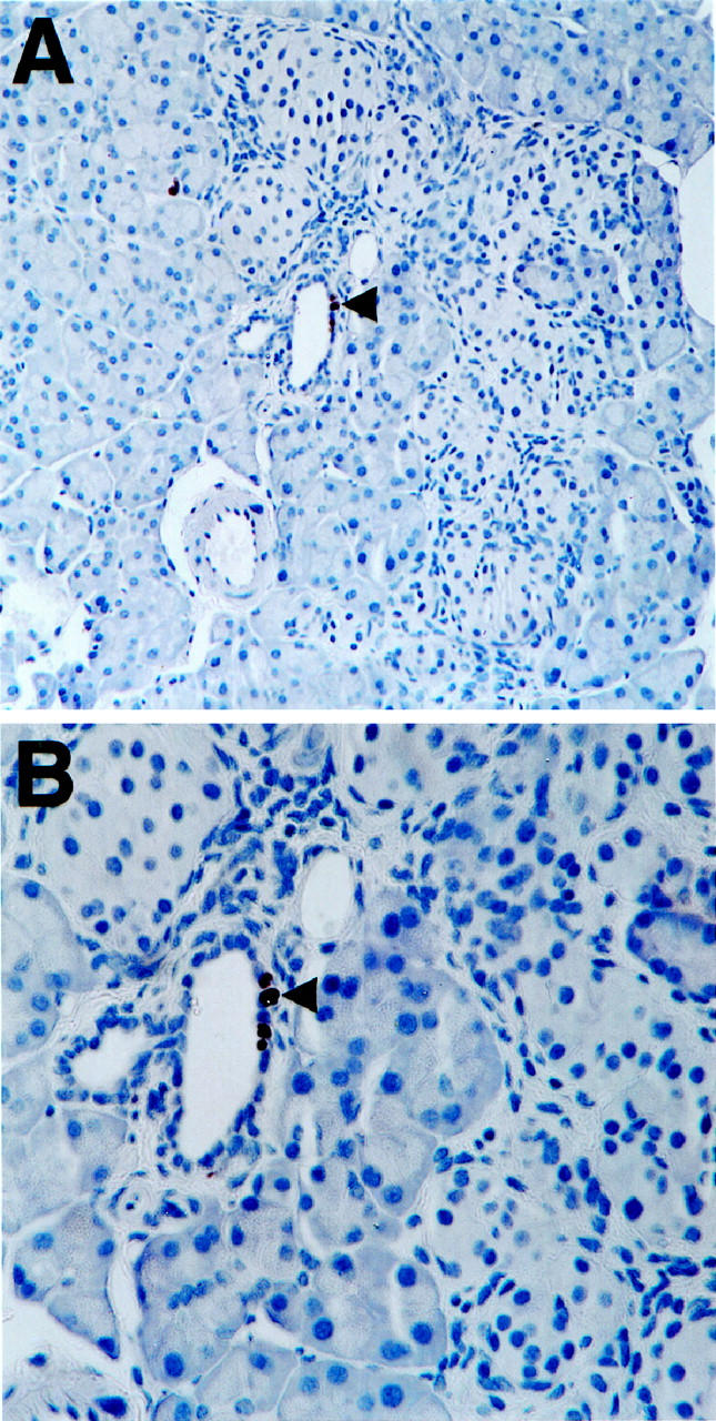
Keratinocyte growth factor, (KGF), a member of the fibroblast growth factor (FGF) family, is involved in wound healing. It also promotes the differentiation of many epithelial tissues and proliferation of epithelial cells as well as pancreatic duct cells. Additionally, many members of the highly homologous FGF family (including KGF), influence both growth and cellular morphology in the developing embryo. We have previously observed elevated levels of KGF in our interferon-gamma transgenic mouse model of pancreatic regeneration. To understand the role of KGF in pancreatic differentiation, we generated insulin promoter-regulated KGF transgenic mice. Remarkably, we have found that ectopic KGF expression resulted in the emergence of hepatocytes within the islets of Langerhans in the pancreas. Additionally, significant intra-islet duct cell proliferation in the pancreata of transgenic KGF mice was observed. The unexpected appearance of hepatocytes and proliferation of intra-islet duct cells in the pancreata of these mice evidently stemmed directly from local exposure to KGF.
Figures
Similar articles
-
Transgenic expression of epidermal growth factor and keratinocyte growth factor in beta-cells results in substantial morphological changes.J Endocrinol. 1999 Aug;162(2):167-75. doi: 10.1677/joe.0.1620167. J Endocrinol. 1999. PMID: 10425454
-
Selective expansion of the beta-cell compartment in the pancreas of keratinocyte growth factor transgenic mice.Am J Physiol Gastrointest Liver Physiol. 2008 May;294(5):G1139-47. doi: 10.1152/ajpgi.00338.2007. Epub 2008 Mar 27. Am J Physiol Gastrointest Liver Physiol. 2008. PMID: 18372394
-
Expression of keratinocyte growth factor in embryonic liver of transgenic mice causes changes in epithelial growth and differentiation resulting in polycystic kidneys and other organ malformations.Oncogene. 1996 May 16;12(10):2109-19. Oncogene. 1996. PMID: 8668336
-
Expression and function of keratinocyte growth factor and activin in skin morphogenesis and cutaneous wound repair.J Investig Dermatol Symp Proc. 2000 Dec;5(1):34-9. doi: 10.1046/j.1087-0024.2000.00009.x. J Investig Dermatol Symp Proc. 2000. PMID: 11147673 Review.
-
Regeneration of pancreatic endocrine cells in interferon-gamma transgenic mice.Adv Exp Med Biol. 1992;321:85-9; discussion 91-3. doi: 10.1007/978-1-4615-3448-8_10. Adv Exp Med Biol. 1992. PMID: 1449085 Review.
Cited by
-
The new stem cell biology: something for everyone.Mol Pathol. 2003 Apr;56(2):86-96. doi: 10.1136/mp.56.2.86. Mol Pathol. 2003. PMID: 12665626 Free PMC article. Review.
-
Direct Lineage Reprogramming: Harnessing Cell Plasticity between Liver and Pancreas.Cold Spring Harb Perspect Biol. 2020 Jul 1;12(7):a035626. doi: 10.1101/cshperspect.a035626. Cold Spring Harb Perspect Biol. 2020. PMID: 31767653 Free PMC article. Review.
-
Disrupted pancreatic exocrine differentiation and malabsorption in response to chronic elevated systemic glucocorticoid.Am J Pathol. 2010 Sep;177(3):1225-32. doi: 10.2353/ajpath.2010.100107. Epub 2010 Jul 22. Am J Pathol. 2010. PMID: 20651242 Free PMC article.
-
Direct cell reprogramming for tissue engineering and regenerative medicine.J Biol Eng. 2019 Feb 13;13:14. doi: 10.1186/s13036-019-0144-9. eCollection 2019. J Biol Eng. 2019. PMID: 30805026 Free PMC article. Review.
-
Recipes for adult stem cell plasticity: fusion cuisine or readymade?J Clin Pathol. 2004 Feb;57(2):113-20. doi: 10.1136/jcp.2003.010074. J Clin Pathol. 2004. PMID: 14747430 Free PMC article. Review.
References
-
- Barr HS: Fibrocystic disease of the pancreas. Lancet 1953, ii:80-83 - PubMed
-
- Porta EA, Stein AA, Patterson P: Ultrastructural changes of the pancreas and liver in cystic fibrosis. Am J Clin Pathol 1964, 42:451-465 - PubMed
-
- Noronha M, Bordalo O, Dreiling DA: Alcohol and the pancreas. II. Pancreatic morphology of advanced alcoholic pancreatitis. Am J Gastroenterol 1981, 76:120-124 - PubMed
-
- Takahashi M, Arai H, Kokubo T, Furukawa F, Kurata Y, Ito N: An ultrastructural study of precancerous and cancerous lesions of the pancreas in Syrian golden hamsters induced by N-nitrosobis(2-oxopropyl)amine. Gann 1981, 71:825-831 - PubMed
Publication types
MeSH terms
Substances
Grants and funding
LinkOut - more resources
Full Text Sources
Other Literature Sources
