Role of macrophage scavenger receptors in hepatic granuloma formation in mice
- PMID: 10079248
- PMCID: PMC1866422
- DOI: 10.1016/S0002-9440(10)65317-5
Role of macrophage scavenger receptors in hepatic granuloma formation in mice
Abstract
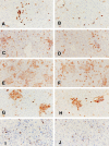
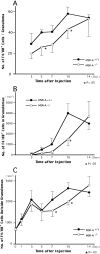
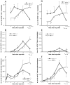
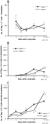
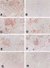
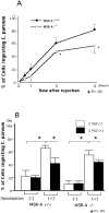
In mice homozygous for the gene mutation for type I and type II macrophage scavenger receptors (MSR-A), MSR-A-/-, the formation of hepatic granulomas caused by a single intravenous injection of heat-killed Corynebacterium parvum was delayed significantly for 10 days after injection, compared with granuloma formation in wild-type (MSR-A+/+) mice. In the early stage of granuloma formation, numbers of macrophages and their precursor cells were significantly reduced in MSR-A-/- mice compared with MSR-A+/+ mice. In contrast to MSR-A+/+ mice, no expression of monocyte chemoattractant protein-1, tumor necrosis factor-alpha, and interferon-gamma mRNA was observed in MSR-A-/- mice by 3 days after injection. Also in MSR-A-/- mice, uptake of C. parvum by Kupffer cells and monocyte-derived macrophages in the early stage of granuloma formation was lower and elimination of C. parvum from the liver was slower than in MSR-A+/+ mice. In the livers of MSR-A+/+ mice, macrophages and sinusoidal endothelial cells possessed MSR-A, but this was not seen in the livers of MSR-A-/- mice. In both MSR-A-/- and MSR-A+/+ mice, expression of other scavenger receptors was demonstrated. These data suggest that MSR-A deficiency impairs the uptake and elimination of C. parvum by macrophages and delays hepatic granuloma formation, particularly in the early stage.
Figures






Similar articles
-
Role of macrophage scavenger receptors in response to Listeria monocytogenes infection in mice.Am J Pathol. 2001 Jan;158(1):179-88. doi: 10.1016/S0002-9440(10)63956-9. Am J Pathol. 2001. PMID: 11141491 Free PMC article.
-
Expression of macrophage colony-stimulating factor and its receptor in hepatic granulomas of Kupffer-cell-depleted mice.Am J Pathol. 1997 Jun;150(6):2047-60. Am J Pathol. 1997. PMID: 9176397 Free PMC article.
-
AIM inhibits apoptosis of T cells and NKT cells in Corynebacterium-induced granuloma formation in mice.Am J Pathol. 2003 Mar;162(3):837-47. doi: 10.1016/S0002-9440(10)63880-1. Am J Pathol. 2003. PMID: 12598318 Free PMC article.
-
Multifunctional roles of macrophages in the development and progression of atherosclerosis in humans and experimental animals.Med Electron Microsc. 2002 Dec;35(4):179-203. doi: 10.1007/s007950200023. Med Electron Microsc. 2002. PMID: 12658354 Review.
-
The multiple roles of macrophage scavenger receptors (MSR) in vivo: resistance to atherosclerosis and susceptibility to infection in MSR knockout mice.J Atheroscler Thromb. 1997;4(1):1-11. doi: 10.5551/jat1994.4.1. J Atheroscler Thromb. 1997. PMID: 9583348 Review.
Cited by
-
Role of macrophage scavenger receptors in response to Listeria monocytogenes infection in mice.Am J Pathol. 2001 Jan;158(1):179-88. doi: 10.1016/S0002-9440(10)63956-9. Am J Pathol. 2001. PMID: 11141491 Free PMC article.
-
Role of granulocyte/macrophage colony-stimulating factor in zymocel-induced hepatic granuloma formation.Am J Pathol. 2001 Jan;158(1):131-45. doi: 10.1016/S0002-9440(10)63951-X. Am J Pathol. 2001. PMID: 11141486 Free PMC article.
-
Pattern recognition scavenger receptors, SR-A and CD36, have an additive role in the development of colitis in mice.Dig Dis Sci. 2009 Dec;54(12):2561-7. doi: 10.1007/s10620-008-0673-4. Dig Dis Sci. 2009. PMID: 19117124 Free PMC article.
-
Receptors, mediators, and mechanisms involved in bacterial sepsis and septic shock.Clin Microbiol Rev. 2003 Jul;16(3):379-414. doi: 10.1128/CMR.16.3.379-414.2003. Clin Microbiol Rev. 2003. PMID: 12857774 Free PMC article. Review.
References
-
- Kodama T, Freeman M, Rohrer L, Zabrecky J, Matsudaira P, Krieger M: Type I macrophage scavenger receptor contains α-helical, and collagen-like coiled coils. Nature 1990, 343:531-535 - PubMed
-
- Rohrer L, Freeman M, Kodama T, Penman M, Krieger M: Coiled-coil fibrous domains mediate ligand binding by macrophage scavenger receptor type II. Nature 1990, 343:570-572 - PubMed
-
- Elomaa O, Kangas M, Sahlberg C, Tuukkanen J, Sormunen S, Liakka A, Thesleff I, Kraal G, Tryggvason K: Cloning of a novel bacteria-binding receptor structurally related to scavenger receptors and expressed in a subset of macrophages. Cell 1995, 80:603-609 - PubMed
-
- van der Laan LJW, Kangas M, Döpp EA, Holub E, Elomaa O, Kraal G, Tryggvason K: Macrophage scavenger receptor MARCO: In vitro and in vivo regulation and involvement in the anti-bacterial host defense. Immunol Lett 1997, 57:203-208 - PubMed
-
- van der Laan LJW, Döpp EA, Haworth R, Pikkarainen T, Kangas M, Elomaa O, Dijkstra CD, Gordon S, Tryggvason K, Kraal G: Regulation and functional involvement of macrophage scavenger receptor MARCO in clearance of bacteria in vivo. J Immunol 1999, 162:939-947 - PubMed
Publication types
MeSH terms
Substances
LinkOut - more resources
Full Text Sources
Other Literature Sources
Medical
Molecular Biology Databases
Research Materials

