

Frequent genetic alterations in simple urothelial hyperplasias of the bladder in patients with papillary urothelial carcinoma
- PMID: 10079249
- PMCID: PMC1866404
- DOI: 10.1016/S0002-9440(10)65318-7
Frequent genetic alterations in simple urothelial hyperplasias of the bladder in patients with papillary urothelial carcinoma
Abstract
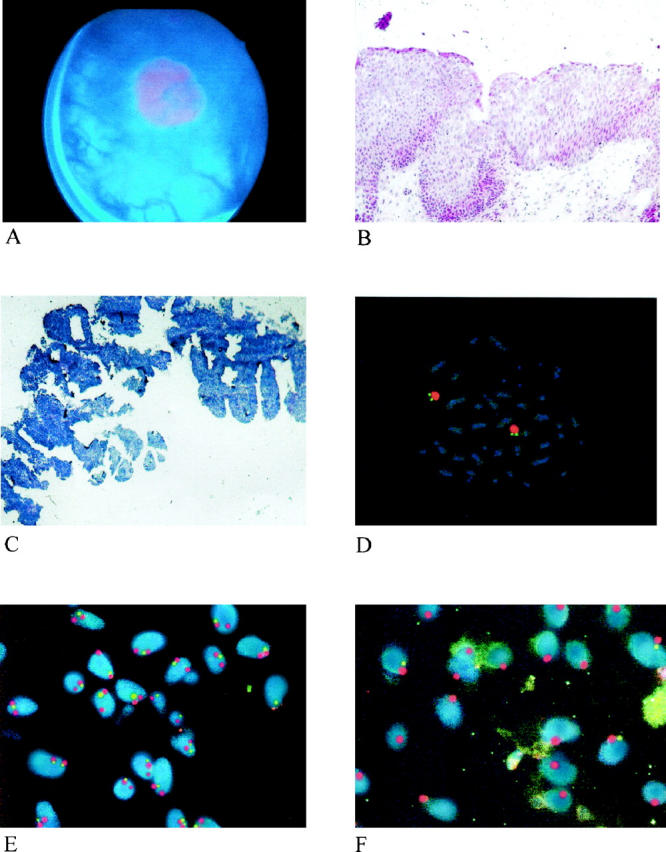
In order to understand the origin of bladder cancer, very early urothelial lesions must be investigated in addition to more advanced tumors. Tissue from 31 biopsies of 12 patients with urothelial hyperplasias and simultaneous or consecutive superficial papillary tumors were used to microdissect urothelium from 15- microm sections of biopsies. The biopsies were obtained with the recently developed highly sensitive diagnostic method of 5-aminolevulinic acid-induced fluorescence endoscopy (AFE). Besides flat and papillary urothelial neoplasms, the method of photodynamic diagnostics also detects simple urothelial hyperplasias as fluorescent positive lesions. In addition, 12 fluorescence-positive biopsies showing histologically normal urothelium were investigated. Fluorescence in situ hybridization was done using a dual color staining technique of biotinylated centromeric probes of chromosomes 9 and 17 and digoxigenin-labeled gene-specific P1 probes for chromosomes 9q22 (FACC), 9p21(p16/CDKI2), and 17p13(p53). Ten of 14 hyperplasias (70%) showed deletions of chromosome 9. In 7 out of 8 patients with genetic alterations in the hyperplasias the genetic change was also present in the papillary tumor. Six out of 12 samples of microdissected normal urothelium also showed genetic alterations on chromosome 9. Microdissection of urothelial lesions, obtained during AFE, has led to the first unequivocal documentation of genetic changes in urothelial lesions diagnosed as normal in histopathology. Thus, this technical approach is important to provide insight into the earliest molecular alterations in bladder carcinogenesis.
Figures
Similar articles
-
Frequent genetic alterations in flat urothelial hyperplasias and concomitant papillary bladder cancer as detected by CGH, LOH, and FISH analyses.J Pathol. 2003 Jan;199(1):50-7. doi: 10.1002/path.1259. J Pathol. 2003. PMID: 12474226
-
Fluorescence in situ hybridization detects frequent chromosome 9 deletions and aneuploidy in histologically normal urothelium of bladder cancer patients.Oncol Rep. 2004 Apr;11(4):745-51. Oncol Rep. 2004. PMID: 15010867
-
Value of multicolour fluorescence in situ hybridisation (UroVysion) in the differential diagnosis of flat urothelial lesions.J Clin Pathol. 2008 Mar;61(3):272-7. doi: 10.1136/jcp.2007.049684. Epub 2007 Aug 10. J Clin Pathol. 2008. PMID: 17693577
-
[Molecular changes in development and progression of urothelial carcinoma].Verh Dtsch Ges Pathol. 2003;87:172-84. Verh Dtsch Ges Pathol. 2003. PMID: 16888910 Review. German.
-
Urothelial dysplasia and other flat lesions of the urinary bladder: clinicopathologic and molecular features.Hum Pathol. 2010 Feb;41(2):155-62. doi: 10.1016/j.humpath.2009.07.002. Epub 2009 Sep 16. Hum Pathol. 2010. PMID: 19762067 Review.
Cited by
-
Expression patterns and prognostic role of transketolase-like 1 in muscle-invasive bladder cancer.World J Urol. 2015 Oct;33(10):1403-9. doi: 10.1007/s00345-014-1473-4. Epub 2015 Jan 9. World J Urol. 2015. PMID: 25572961
-
Comparison of the clonality of urothelial carcinoma developing in the upper urinary tract and those developing in the bladder.Springerplus. 2013 Aug 28;2:412. doi: 10.1186/2193-1801-2-412. eCollection 2013. Springerplus. 2013. PMID: 24024098 Free PMC article.
-
5'-S-(3-Aminophenyl)-5'-thioadenosine, a Novel Chemoprotective Agent for Reducing Toxic Side Effects of Fluorouracil in Treatment of MTAP-Deficient Cancers.Mol Cancer Ther. 2025 Jul 2;24(7):1030-1039. doi: 10.1158/1535-7163.MCT-24-0656. Mol Cancer Ther. 2025. PMID: 40062378
-
Photodynamic Diagnosis and Therapy in Non-Muscle-Invasive Bladder Cancer.Cancers (Basel). 2024 Jun 22;16(13):2299. doi: 10.3390/cancers16132299. Cancers (Basel). 2024. PMID: 39001362 Free PMC article. Review.
-
Biomarkers of Bladder Cancer: Cell-Free DNA, Epigenetic Modifications and Non-Coding RNAs.Int J Mol Sci. 2022 Oct 30;23(21):13206. doi: 10.3390/ijms232113206. Int J Mol Sci. 2022. PMID: 36361996 Free PMC article. Review.
References
-
- Amin MB, Young RH: Intraepithelial lesions of the urinary bladder with a discussion of the histogenesis of urothelial neoplasia. Semin Diagn Pathol 1997, 14:84-97 - PubMed
-
- Kriegmair M, Baumgartner R, Knuchel R, Stepp H, Hofstadter F, Hofstetter A: Detection of early bladder cancer by 5-aminolevulinic acid induced porphyrin fluorescence [see comments]. J Urol 1996, 155:105-109 - PubMed
-
- Steinbach P, Kriegmair M, Baumgartner R, Hofstadter F, Knuchel R: Intravesical instillation of 5-aminolevulinic acid: the fluorescent metabolite is limited to urothelial cells. Urology 1994, 44:676-668 - PubMed
-
- Peng Q, Warloe T, Berg K, Moan J, Kongshaug M, Giercksky KE, Nesland JM: 5-Aminolevulinic acid-based photodynamic therapy: clinical research and future challenges. Cancer 1997, 79:2282-2308 - PubMed
-
- Chatuverdi V, Hodges S, Johnston D, Ro JY, Logothetis C, von Eschenbach AC, Batsakis JG, Czerniak B: Superimposed histologic and genetic mapping of chromosome 17 alterations in human urinary bladder neoplasia. Oncogene 1997, 14:2059-2070 - PubMed
Publication types
MeSH terms
LinkOut - more resources
Full Text Sources
Medical
Research Materials
Miscellaneous
