. 1999 Mar;154(3):871-82.
doi: 10.1016/s0002-9440(10)65334-5.

The compliance of collagen gels regulates transforming growth factor-beta induction of alpha-smooth muscle actin in fibroblasts

Affiliations

The compliance of collagen gels regulates transforming growth factor-beta induction of alpha-smooth muscle actin in fibroblasts

P D Arora et al. Am J Pathol. 1999 Mar.

Abstract

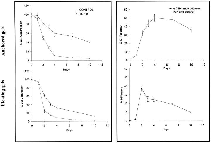
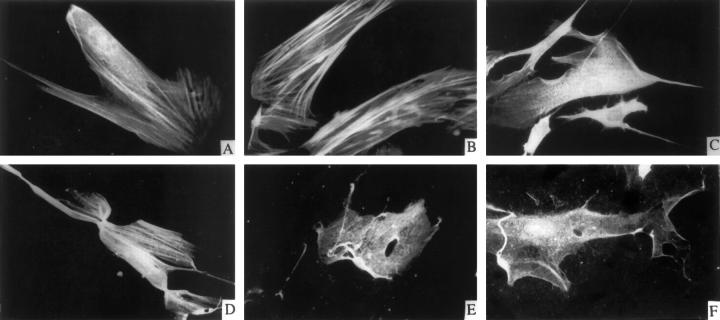
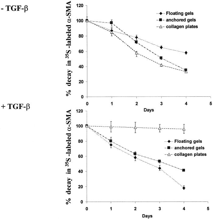
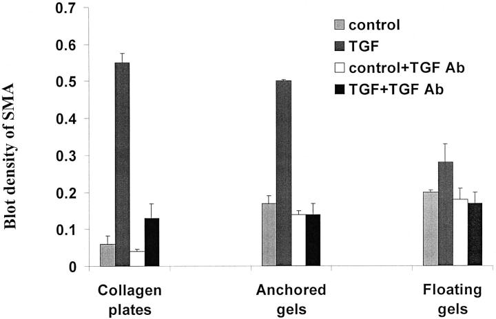
Wound contraction is mediated by myofibroblasts, specialized fibroblasts that appear in large numbers as the wound matures and when resistance to contractile forces increases. We considered that the regulation of myofibroblast differentiation by wound-healing cytokines may be dependent on the resistance of the connective tissue matrix to deformation. We examined transforming growth factor-beta1 (TGF-beta1) induction of the putative fibroblast contractile marker, alpha-smooth muscle actin (alpha-SMA), and the regulation of this process by the compliance of collagen substrates. Cells were cultured in three different types of collagen gels with wide variations of mechanical compliance as assessed by deformation testing. The resistance to collagen gel deformation determined the levels of intracellular tension as shown by staining for actin stress fibers. For cells plated on thin films of collagen-coated plastic (ie, minimal compliance and maximal intracellular tension), TGF-beta1 (10 ng/ml; 6 days) increased alpha-SMA protein content by ninefold as detected by Western blots but did not affect beta-actin content. Western blots of cells in anchored collagen gels (moderate compliance and tension) also showed a TGF-beta1-induced increase of alpha-SMA content, but the effect was greatly reduced compared with collagen-coated plastic (<3-fold increase). In floating collagen gels (high compliance and low tension), there were only minimal differences of alpha-SMA protein. Northern analyses for alpha-SMA and beta-actin indicated that TGF-beta1 selectively increased mRNA for alpha-SMA similar to the reported protein levels. In pulse-chase experiments, [35S]methionine-labeled intracellular alpha-SMA decayed most rapidly in floating gels, less rapidly in anchored gels, and not at all in collagen plates after TGF-beta1 treatment. TGF-beta1 increased alpha2 and beta1 integrin content by 50% in cells on collagen plates, but the increase was less marked on anchored gels and was undetectable in floating gels. When intracellular tension on collagen substrates was reduced by preincubating cells with blocking antibodies to the alpha2 and beta1 integrin subunits, TGF-beta1 failed to increase alpha-SMA protein content in all three types of collagen matrices. These data indicate that TGF-beta1-induced increases of alpha-SMA content are dependent on the resistance of the substrate to deformation and that the generation of intracellular tension is a central determinant of contractile cytoskeletal gene expression.


Figures

Figure 1.
Figure 1.
Collagen gel contraction and regulation by TGF-β1. Left panels: Cells in anchored (top) and floating (bottom) collagen gels were incubated in α-MEM containing 3H2O (1 μCi/ml) with or without TGF-β1 (10 ng/ml). At 1, 2, 3, 4, 7, and 10 days, gel volume was determined using scintillation counting of labeled gels. For each time point, n = 3 independent samples. Results are presented as mean percent gel volume ± SEM. Right panels: Data from left panels re-plotted to show percent differences between TGF-β1-treated cultures and controls.
Figure 2.
Figure 2.
Immunofluorescence of α-SMA in cells on different substrates. Confocal micrographs of cells on collagen-coated plastic (A and B), anchored collagen gels (C and D), and floating collagen gels (E and F) were incubated with vehicle (A, C, and E) or with TGF-β1 (10 ng/ml; B, D, and F) for 3 days. α-SMA was immunostained with anti-α-SMA antibody and FITC-conjugated goat anti-mouse antibody.
Figure 3.
Figure 3.
Western blots of α-SMA protein content. A: Cells on plastic plates were incubated with vehicle control or TGF-β1 (10 ng/ml) for 3 days. Immunoblots of α-SMA and β-actin were scanned and the densities plotted (mean density ± SEM). B: Cells were preincubated for 3 days with vehicle or with TGF-β1 (10 ng/ml) on plastic plates as in A and then inoculated on collagen-coated plates or anchored collagen gels or in floating gels and incubated again with or without 10 ng/ml TGF-β1 for another 3 days. C: Both control and experimental cells were preincubated with TGF-β1 (10 ng/ml) for 3 days and then inoculated on collagen-coated plates or anchored or floating collagen gels for 3 days and treated either with vehicle (controls) or with TGF-β1. Western blotting and densitometry were used to assess α-SMA content and β-actin content in three independent samples. Data are expressed as a ratio of the blot densities of α-SMA to β-actin (mean ratio ± SEM).
Figure 4.
Figure 4.
Northern blots of α-SMA and β-actin. Cells were prepared as in Figure 3 ▶ . A: Fibroblasts were incubated with TGF-β1 or vehicle on tissue culture plastic for 3 days, and mRNA levels were measured by Northern hybridization using oligonucleotide probes specific for either human α-SMA or β-actin. B: Cells were preincubated with either vehicle or TGF-β as in A and then inoculated in collagen-coated plates, anchored collagen gels, or floating collagen gels and treated with or without TGF-β1 for 3 days. Cells were assayed for α-SMA and β-actin mRNAs by Northern hybridization. Note the lack of up-regulation of α-SMA by TGF-β1 in floating gels.
Figure 5.
Figure 5.
Loss of nascent α-SMA by metabolic labeling analyses. Cells were preincubated with vehicle or TGF-β and labeled for the last 24 hours on plastic plates with [35S]methionine followed by inoculation of cells in different types of collagen gels for up to 4 days with or without TGF-β as indicated. Cells were released from gels and subjected to immunoprecipitation with anti-α-SMA antibody. The relative amounts of nascent α-SMA content were estimated from fluorographic blot densities and expressed as percentage of starting material (ie, day 0 of incubation in gels). Results are expressed as mean ± SEM percent starting material.
Figure 6.
Figure 6.
TGF-β antibody blockade of α-SMA induction. Cells were preincubated with vehicle or TGF-β1 for 3 days on plastic plates as in Figure 3 ▶ , inoculated into the three different types of collagen gels and incubated with PBS (control) or TGF-β1 (TGF) with or without 15 μg/ml anti-TGF-β neutralizing antibody (TGF Ab) for 3 days. Three independent samples were analyzed for the expression of α-SMA by Western blot. Results are expressed as mean density of α-SMA ± SEM. Values are normalized to β-actin densities for each lane.
Figure 7.
Figure 7.
Integrin content of cells in collagen gels. Cells grown on tissue culture plastic were preincubated with vehicle or TGF-β and then inoculated into different types of collagen gels. Cells were incubated with or without TGF-β1 for another 3 days. Three independent samples were analyzed by Western blot for α2-integrin or β1-integrin by Western blots. β-actin was used for normalization of protein loading for each lane.
Figure 8.
Figure 8.
A to E:Phase contrast photomicrographs of cells treated with α2- and/or β1-integrin inhibiting antibodies. Cells were incubated with vehicle control (A), TGF-β1 alone (B), TGF-β1 with anti-α2-integrin antibody (C), anti-β1-integrin antibody (D), or both anti-integrin antibodies (E). Micrographs were obtained on day 3 of treatment on collagen-coated plates. Magnification, ×150. F: Fibroblasts on collagen-coated plates, anchored gels, or floating gels were incubated with either no antibody (control) or with only anti-α2-integrin antibody or with only anti-β1-integrin in the presence or absence of TGF-β1 for 3 days. Antibodies were replenished once during the incubation period. Three independent samples were assayed by Western blot for expression of α-SMA and normalized against β-actin. Results are expressed as mean density ± SEM.

Similar articles

Cited by

References

    1. Skalli O, Gabbiani G: The biology of the myofibroblast. Molecular and Cellular Biology of Wound Repair. Edited by Clark RAF, Henson PM. New York, Plenum, 1996
    1. Darby I, Skalli O, Gabbiani G: α-Smooth muscle actin is transiently expressed by myofibroblasts during experimental wound healing. Lab Invest 1990, 63:21-29 - PubMed
    1. Singer II, Kawka DW, Kazazis DM, Clark RAF: The in vivo codistribution of fibronectin and actin fibers in granulation tissue: immunofluorescence and electron microscopic studies of the fibronexus at the myofibroblast surface. J Cell Biol 1984, 98:2091-2106 - PMC - PubMed
    1. Burridge K: Are stress fibers contractile? Nature 1981, 249:61-62 - PubMed
    1. Gabbiani G, Ryan GB, Majno G: Presence of modified fibroblasts in granulation tissue and their possible role in wound contraction. Experientia 1971, 27:549-550 - PubMed

Publication types