

Helicobacter pylori-induced chronic active gastritis, intestinal metaplasia, and gastric ulcer in Mongolian gerbils
- PMID: 10079274
- PMCID: PMC1866409
- DOI: 10.1016/S0002-9440(10)65343-6
Helicobacter pylori-induced chronic active gastritis, intestinal metaplasia, and gastric ulcer in Mongolian gerbils
Abstract
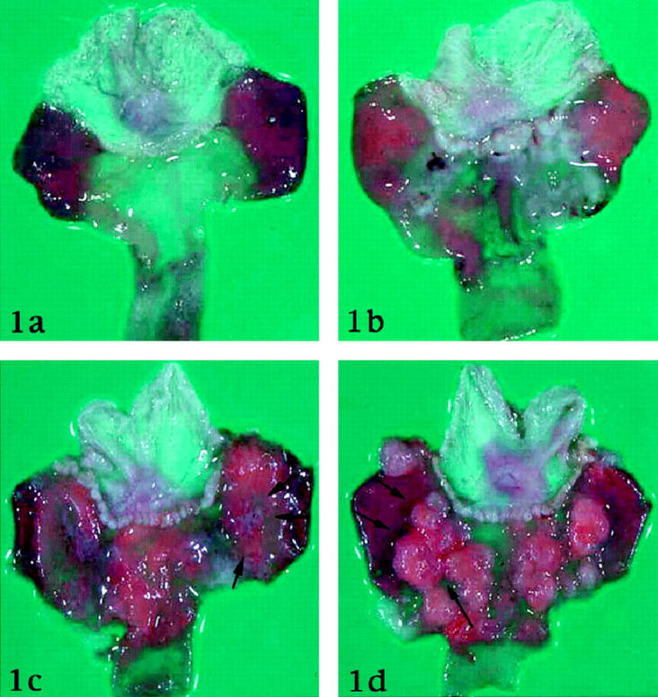
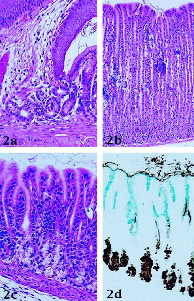
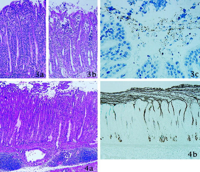
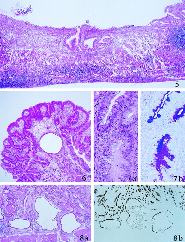
The establishment of persisting Helicobacter pylori infection in laboratory animals has been difficult, but in 1996 Hirayama reported the development of a successful Mongolian gerbil model. The present study was undertaken with two aims: to better characterize the normal histological structure and histochemical properties of the gastric mucosa of the Mongolian gerbil; and to evaluate the progression of the histopathological features of H. pylori-induced gastritis in this animal model for one year after the experimental infection. Seventy-five Mongolian gerbils were used. Mongolian gerbils were sacrificed at 2, 4, 8, 12, 26, 38, and 52 weeks after H. pylori inoculation. Sections prepared from stomachs immediately fixed in Carnoy's solution were stained with hematoxylin and eosin and Alcian blue at pH 2.5/periodic acid-Schiff, a dual staining consisting of the galactose oxidase-cold thionin Schiff reaction and paradoxical Concanavalin A staining, and with immunostaining for H. pylori and BrdU. H. pylori infection induced in the Mongolian gerbil a chronic active gastritis, in which a marked mucosal infiltration of neutrophils on a background of chronic inflammation became detectable 4 weeks after inoculation and continued up to 52 weeks. Intestinal metaplasia and gastric ulcers appeared after 26 weeks in some of the animals, whereas others developed multiple hyperplastic polyps. The Mongolian gerbil represents a novel and useful model for the study of H. pylori-induced chronic active gastritis and may lend itself to the investigation of the epithelial alterations that lead to intestinal metaplasia and gastric neoplasia.
Figures
Similar articles
-
Long-term treatment with sterigmatocystin, a fungus toxin, enhances the development of intestinal metaplasia of gastric mucosa in Helicobacter pylori-infected Mongolian gerbils.Scand J Gastroenterol. 2003 Apr;38(4):360-9. Scand J Gastroenterol. 2003. PMID: 12739707
-
Pathological changes in glandular stomach of Helicobacter pylori-infected Mongolian gerbil model.J Gastroenterol. 1998;33 Suppl 10:22-5. J Gastroenterol. 1998. PMID: 9840012
-
Development of gastric adenocarcinoma in Mongolian gerbils after long-term infection with Helicobacter pylori.J Gastroenterol Hepatol. 2004 Oct;19(10):1192-8. doi: 10.1111/j.1440-1746.2004.03469.x. J Gastroenterol Hepatol. 2004. PMID: 15377299
-
[Pharmacological study on the pathological changes of the gastric mucosa in Helicobacter pylori-infected Mongolian gerbils].Nihon Yakurigaku Zasshi. 2001 Oct;118(4):259-68. doi: 10.1254/fpj.118.259. Nihon Yakurigaku Zasshi. 2001. PMID: 11680169 Review. Japanese.
-
[Rodent models of Helicobacter pylori infection and their utility].Nihon Yakurigaku Zasshi. 1998 May;111(5):289-96. doi: 10.1254/fpj.111.289. Nihon Yakurigaku Zasshi. 1998. PMID: 9666483 Review. Japanese.
Cited by
-
Standardized ginger (Zingiber officinale) extract reduces bacterial load and suppresses acute and chronic inflammation in Mongolian gerbils infected with cagAHelicobacter pylori.Pharm Biol. 2009;47(1):92-98. doi: 10.1080/13880200802448690. Pharm Biol. 2009. PMID: 20376296 Free PMC article.
-
Rapid loss of motility of Helicobacter pylori in the gastric lumen in vivo.Infect Immun. 2005 Mar;73(3):1584-9. doi: 10.1128/IAI.73.3.1584-1589.2005. Infect Immun. 2005. PMID: 15731057 Free PMC article.
-
Gastric mucosal interleukin-17 and -18 mRNA expression in Helicobacter pylori-induced Mongolian gerbils.Cancer Sci. 2009 Nov;100(11):2152-9. doi: 10.1111/j.1349-7006.2009.01291.x. Epub 2009 Jul 21. Cancer Sci. 2009. PMID: 19694753 Free PMC article.
-
Severity of gastritis determines glandular stomach carcinogenesis in Helicobacter pylori-infected Mongolian gerbils.Cancer Sci. 2007 Apr;98(4):478-83. doi: 10.1111/j.1349-7006.2007.00416.x. Epub 2007 Jan 31. Cancer Sci. 2007. PMID: 17284248 Free PMC article.
-
Characterization of virulence factors of mouse-adapted Helicobacter pylori strain SS1 and effects on gastric hydrophobicity.Dig Dis Sci. 2001 Sep;46(9):1943-51. doi: 10.1023/a:1010691216207. Dig Dis Sci. 2001. PMID: 11575447
References
-
- Karita M, Kouchiyama T, Okita K, Nakazawa T: New small animal model for human gastric Helicobacter pylori infection: success in both nude and euthymic mice. Am J Gastroenterol 1991, 86:1596-1603 - PubMed
-
- Karita M, Li Q, Cantero D, Okita K: Establishment of a small animal model for human Helicobacter pylori infection using germ-free mouse. Am J Gastroenterol 1994, 89:208-213 - PubMed
-
- Lee A, O’Rourke J, De Ungria MC, Robertson B, Daskalopoulos G, Dixon MF: A standardized mouse model of Helicobacter pylori infection: introducing the Sydney strain. Gastroenterology 1997, 112:1386-1397 - PubMed
MeSH terms
LinkOut - more resources
Full Text Sources
Medical
