Comparative Study
. 1999 Apr;19(4):2535-46.
doi: 10.1128/MCB.19.4.2535.

The schizosaccharomyces pombe dim1(+) gene interacts with the anaphase-promoting complex or cyclosome (APC/C) component lid1(+) and is required for APC/C function

Affiliations
Comparative Study

The schizosaccharomyces pombe dim1(+) gene interacts with the anaphase-promoting complex or cyclosome (APC/C) component lid1(+) and is required for APC/C function

L D Berry et al. Mol Cell Biol. 1999 Apr.

Abstract

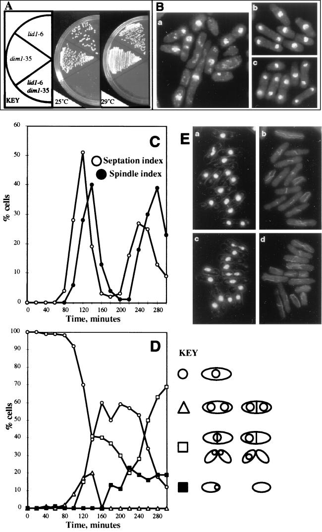
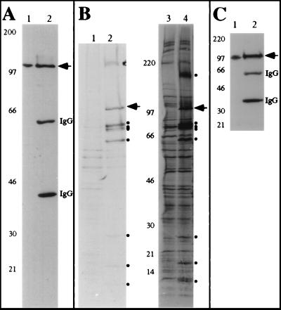
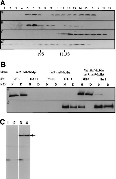
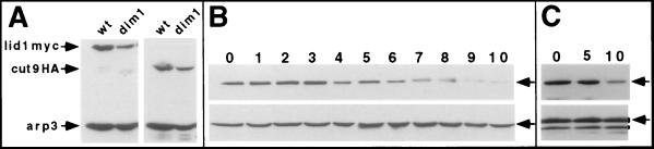
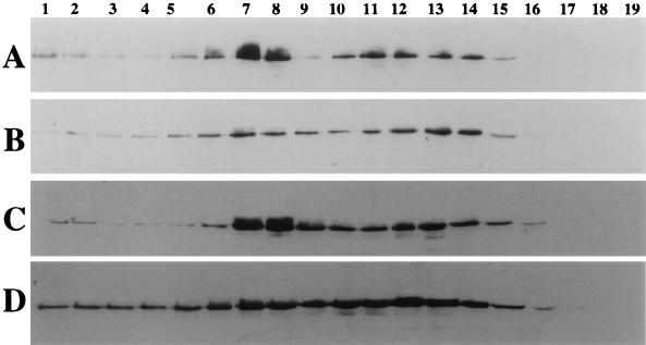
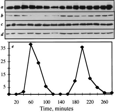
The Schizosaccharomyces pombe dim1(+) gene is required for entry into mitosis and for chromosome segregation during mitosis. To further understand dim1p function, we undertook a synthetic lethal screen with the temperature-sensitive dim1-35 mutant and isolated lid (for lethal in dim1-35) mutants. Here, we describe the temperature-sensitive lid1-6 mutant. At the restrictive temperature of 36 degrees C, lid1-6 mutant cells arrest with a "cut" phenotype similar to that of cut4 and cut9 mutants. An epitope-tagged version of lid1p is a component of a multiprotein approximately 20S complex; the presence of lid1p in this complex depends upon functional cut9(+). lid1p-myc coimmunoprecipitates with several other proteins, including cut9p and nuc2p, and the presence of cut9p in a 20S complex depends upon the activity of lid1(+). Further, lid1(+) function is required for the multiubiquitination of cut2p, an anaphase-promoting complex or cyclosome (APC/C) target. Thus, lid1p is a component of the S. pombe APC/C. In dim1 mutants, the abundances of lid1p and the APC/C complex decline significantly, and the ubiquitination of an APC/C target is abolished. These data suggest that at least one role of dim1p is to maintain or establish the steady-state level of the APC/C.


Figures

FIG. 1
FIG. 1
lid1-6 mutant phenotype. (A) lid1-6 and dim1-35 display a near-lethal synthetic interaction. lid1-6, dim1-35, or lid1-6 dim1-35 cells were streaked to YE agar and then incubated at 25 or 29°C for 3 days. (B) lid1-6 dim1-35 (panel a), lid1-6 (panel b), or dim1-35 (panel c) cells were grown at 25°C, fixed, and stained with DAPI. (C to E) A synchronous population of lid1-6 cells shifted to a restrictive temperature fails to undergo spindle elongation or chromosome segregation at the first mitosis. Subsequent septation results in missegregation of DNA or development of a cut phenotype. Cells were synchronized in early G2 by centrifugal elutriation and then inoculated into rich medium at 36°C. Samples were collected at 20-min intervals and subjected to various analyses. (C) Spindle index and septation index of lid1-6 synchronous cells after the shift. (D) Phenotypes of lid1-6 cells after the shift. Note that at the first mitosis, a small percentage of cells undergo a normal anaphase (▵), as evidenced by normal DNA segregation observed by DAPI staining. In the majority of cells, however, normal anaphase is not observed. Rather, cells cut or missegregate their DNA (■ and □). (E) DAPI (panel a) and tubulin (panel b) staining at 120 min after the shift (corresponding to the first spindle index peak) (note the abundance of short mitotic spindles and the absence of elongated spindles or normally segregated DNA) and DAPI (panel c) and tubulin (panel d) staining at 140 min after the shift (corresponding to the first peak of septation) (note that the majority of septated cells have cut or missegregated their DNA).
FIG. 2
FIG. 2
Cloning of lid1+. (A) Restriction map of the lid1+ genomic locus. Shown below the restriction map is the lid1+ coding region. ■, intron; *, stop codon. Shown at bottom is the deletion construct. (B) DNA sequence and predicted amino acid sequence of lid1+. Intron sequences are in lowercase letters. *, stop codon.
FIG. 2
FIG. 2
Cloning of lid1+. (A) Restriction map of the lid1+ genomic locus. Shown below the restriction map is the lid1+ coding region. ■, intron; *, stop codon. Shown at bottom is the deletion construct. (B) DNA sequence and predicted amino acid sequence of lid1+. Intron sequences are in lowercase letters. *, stop codon.
FIG. 3
FIG. 3
lid1p-myc associates with several other proteins in vivo. (A) lid1p-myc is an ∼100-kDa protein. KGY1302 whole-cell lysates (lane 1) or 9E10 immunoprecipitates from KGY1302 lysates (lane 2) were resolved by SDS-PAGE and transferred to a PVDF membrane, and the membrane was probed with the 9E10 antibody. Molecular mass standards (in kilodaltons) are indicated. (B) Wild-type (lanes 1 and 3) or KGY1302 (lid1::lid1-myc) (lanes 2 and 4) native 35S-labeled cell lysates were immunoprecipitated with the 9E10 antibody, and immunoprecipitates were resolved by SDS-PAGE. Labeled proteins from two separate experiments for 1 day (lanes 1 and 2) or 7 days (lanes 3 and 4) were visualized by fluorography. Migrations of molecular mass markers (in kilodaltons) are indicated. Bands specific to the KGY1302 immunoprecipitate are indicated (●). An arrowhead indicates the band corresponding to lid1p-myc. (C) Cut9p-HA is an ∼90-kDa protein. A KGY1365 whole-cell lysate (lane 1) or an HA.11 immunoprecipitate (lane 2) was resolved by SDS-PAGE and transferred to a PVDF membrane, and the membrane was probed with the HA.11 antibody.
FIG. 4
FIG. 4
lid1p-myc is a component of the 20S APC/C. (A) lid1p-myc and cut9p-HA cosediment at ∼20S. Fractions were collected from the bottom (fraction 1) of 10 to 30% sucrose gradients and then resolved by SDS-PAGE and immunoblotted with the 9E10 antibody to detect lid1p-myc (a and c) or with 12CA5 to detect cut9-HA (b and d). Panels a and b show identical fractions from lysates prepared from the lid1::lid1-myc cut9::cut9::3xHA strain. Panel c shows fractions from the lid1::lid1-myc cut9-665 strain that had been shifted to 36°C for 4 h prior to lysis. Panel d shows fractions from the cut9::cut9-HA lid1-6 strain that had been shifted to 36°C for 4 h prior to lysis. Fraction numbers are indicated above the panels. Peak fractions of the 19S sedimentation marker thyroglobulin and the 11.3S sedimentation marker catalase are indicated below the panels. (B) lid1-myc and cut9p-HA coimmunoprecipitate from KGY1366 (lid1::lid1-myc cut9::cut9-HA) whole-cell lysates but not from KGY1302 (lid1::lid1-myc) or KGY1365 (cut9::cut9-HA) cell lysates. KGY1302 (lanes 1, 2, 3, and 4), KGY1365 (lanes 5, 6, 7, and 8), or KGY1366 (lanes 9, 10, 11, and 12) native (N) (lanes 1, 3, 5, 7, 9, and 11) or denatured (D) (lanes 2, 4, 6, 8, 10, and 12) cell lysates were immunoprecipitated (IP) with 9E10 (lanes 1, 2, 5, 6, 9, and 10) or HA.11 (lanes 3, 4, 7, 8, 11, and 12) antibodies. Immunoprecipitates were resolved by SDS-PAGE and then subjected to immunoblotting with 9E10 (a) or HA.11 (b) antibodies. (C) lid1p-myc coimmunoprecipitates with cut9p and nuc2p. Polyclonal antibodies to cut9p (lanes 1 and 3) or nuc2p (lanes 2 or 4) were used for immunoprecipitation from wild-type (lanes 1 and 2) or KGY1302 (lanes 3 and 4) cell lysates. The immunoprecipitates were resolved by SDS-PAGE and blotted with 9E10 antibodies. lid1p-myc is indicated with an arrow.
FIG. 5
FIG. 5
lid1+ and dim1+ functions are required for ubiquitination of cut2p. (A) Total cell lysates from wild-type (lane 1) or cut2::cut2-myc (lane 2) cells were resolved by SDS-PAGE and immunoblotted with 9E10. In the cut2p-myc doublet in lane 2, the lower-molecular mass protein is likely to be a degradation product of cut2p-myc. (B) His6-ubiquitin was expressed from the nmt1 promoter for 22 h at 25°C in wild-type cells (lane 1), mts3-1 mutant cells (lane 2), mts3-1 cut::cut2-myc lid1-6 cells (lane 3), or mts3-1 cut::cut2-myc cells (lane 4), and the strains were then shifted to 36°C for a further 4 h. His6-ubiquitin conjugates were purified on Ni2+-NTA columns as described in Materials and Methods, and ubiquitinated cut2p-myc was detected by immunoblotting with 9E10. (C) As in panel B except that the strains examined were the wild-type (lane 1), mts3-1 (lane 2), mts3-1 cut::cut2-myc (lanes 3 and 5), mts3-1 cut::cut2-myc lid1-6 (lane 4), and mts3-1 cut::cut2-myc dim1-35 (lane 6) strains. (D) Prior to purification on Ni2+-NTA columns, an aliquot of each lysate used for panel C was examined for the presence of cut2p-myc by immunoblotting with 9E10. Lysates were from wild-type cells (lane 1), mts3-1 mutant cells (lane 2), mts3-1 cut::cut2-myc cells (lane 3), mts3-1 cut::cut2-myc dim1-35 cells (lane 4), and mts3-1 cut::cut2-myc lid1-6 cells (lane 5). The arrow indicates the position of cut2p-myc. Numbers on the left in panels A to C are molecular masses in kilodaltons.
FIG. 6
FIG. 6
lid1p-myc levels drop in the absence of dim1+ function. (A) Wild-type (wt) or dim1-35 (dim1) cells producing lid1p-myc or cut9p-HA were grown to mid-log phase at 25°C and then shifted to 36°C for 4 h. Total protein lysates were prepared and immunoblotted with 9E10 (left panel) or 12CA5 (right panel) antibodies. Immunoblotting with anti-arp3p antibodies provided a loading control. (B and C) The dim1::his3+ strain carrying a single integrated copy of nmt1-T81::dim1+ (KGY1180) and the lid1::lid1-myc cassette was grown in minimal medium lacking thiamine and then shifted to rich medium containing thiamine to repress expression of the nmt1-T81::dim1+ cassette. Samples were collected for analysis at the indicated times (given in hours). Protein lysates were prepared and immunoblotted for lid1p-myc (top panels), arp3p (bottom panel in panel B), or cdc2p with anti-PSTAIRE monoclonal antibody (bottom panel in panel C).
FIG. 7
FIG. 7
dim1+ function affects the abundance of the 20S APC/C. cut9::cut9-HA (A and C) and cut9::cut9-HA dim1-35 (B and D) cells were grown to mid-log phase at 25°C and shifted to 36°C for 4 h (A and B) or 6 h (C and D). Cell lysates were prepared and layered over sucrose gradients. Fractions were collected from the bottom (fraction 1), resolved by SDS-PAGE, and immunoblotted with 12CA5 monoclonal antibodies. Reactive proteins were visualized by ECL. Fraction numbers are indicated above the panels. Peak fractions of the 19S sedimentation marker thyroglobulin and the 11.3S sedimentation marker catalase are indicated below the panels.
FIG. 8
FIG. 8
lid1p-myc levels remain constant through the cell cycle. KGY1366 cells were synchronized in early G2 by centrifugal elutriation and then released into fresh medium. As cells progressed through the cell cycle, synchrony was monitored by determination of septation index at 20-min intervals (e). At the same intervals, samples were collected for analysis of cut9p-HA (a), cdc13p (b), lid1p-myc (c), and arp3p (d) protein levels.
FIG. 9
FIG. 9
Sequence comparison of lid1p with human APC4 and S. cerevisiae Apc4p. The indicated amino acid sequences of human APC4 (top line), S. pombe lid1p (middle line), and S. cerevisiae Apc4p (bottom line) were aligned. Amino acids identical in lid1p and APC4 or Apc4p are indicated by vertical dashes. Amino acids identical in APC4 and Apc4p are indicated by asterisks.

References

    1. Bahler J, Wu J Q, Longtine M S, Shah N G, McKenzie III A, Steever A B, Wach A, Philippsen P, Pringle J R. Heterologous modules for efficient and versatile PCR-based gene targeting in Schizosaccharomyces pombe. Yeast. 1998;14:943–951. - PubMed
    1. Balasubramanian M K, McCollum D, Gould K L. Cytokinesis in fission yeast Schizosaccharomyces pombe. Methods Enzymol. 1997;283:494–506. - PubMed
    1. Barbet N, Muriel W J, Carr A M. Versatile shuttle vectors and genomic libraries for use with Schizosaccharomyces pombe. Gene. 1992;114:59–66. - PubMed
    1. Benito J, Martin-Castellanos C, Moreno S. Regulation of the G1 phase of the cell cycle by periodic stabilization and degradation of the p25rum1 CDK inhibitor. EMBO J. 1998;17:482–497. - PMC - PubMed
    1. Berry L D, Gould K L. Fission yeast dim1(+) encodes a functionally conserved polypeptide essential for mitosis. J Cell Biol. 1997;137:1337–1354. - PMC - PubMed

Publication types

MeSH terms

LinkOut - more resources