. 1999 Apr;19(4):2712-23.
doi: 10.1128/MCB.19.4.2712.

Reciprocal interaction between two cellular proteins, Puralpha and YB-1, modulates transcriptional activity of JCVCY in glial cells

Affiliations

Reciprocal interaction between two cellular proteins, Puralpha and YB-1, modulates transcriptional activity of JCVCY in glial cells

M Safak et al. Mol Cell Biol. 1999 Apr.

Abstract

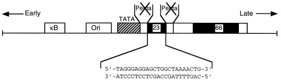
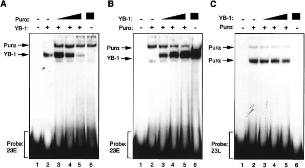
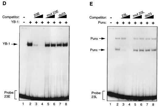
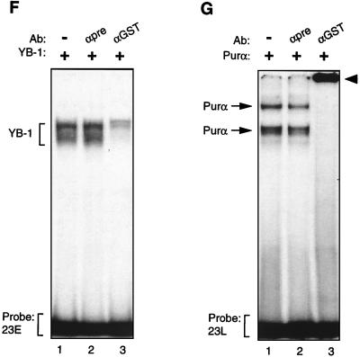
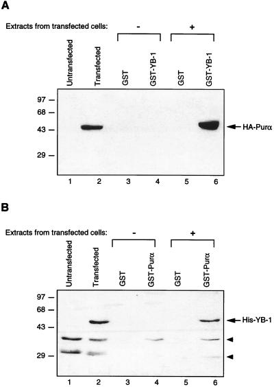
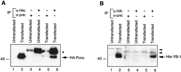
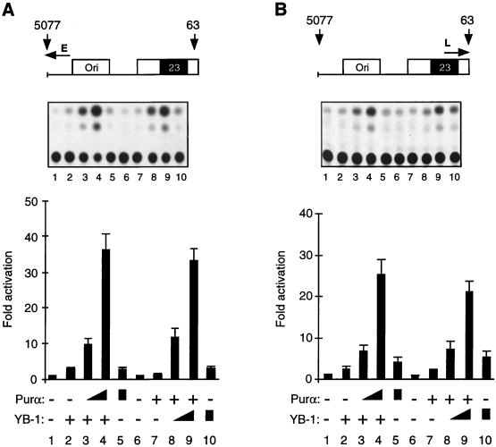
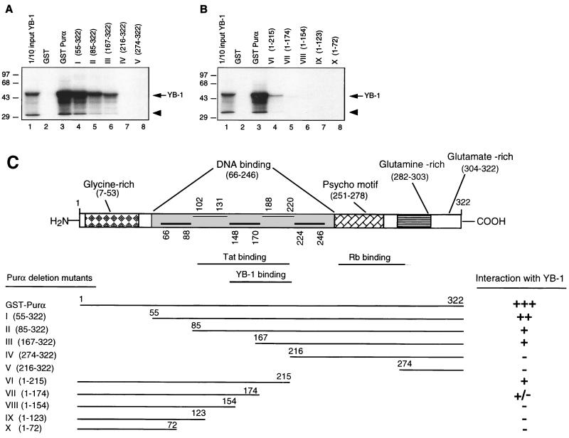
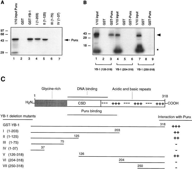
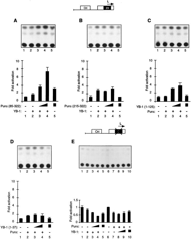
Cross communication between regulatory proteins is an important event in the control of eukaryotic gene transcription. Here we have examined the structural and functional interaction between two cellular regulatory proteins, YB-1 and Puralpha, on the 23-bp sequence element derived from the enhancer-promoter of the human polyomavirus JCV. YB-1 and Puralpha are single-stranded DNA binding proteins which recognize C/T- and GC/GA-rich sequences, respectively. Results from band shift studies demonstrated that while both proteins interact directly with their DNA target sequences within the 23-bp motif, each protein can regulate the association of the other one with the DNA. Affinity chromatography and coimmunoprecipitation provide evidence for a direct interaction between Puralpha and YB-1 in the absence of the DNA sequence. Ectopic expression of YB-1 and Puralpha in glial cells synergistically stimulated viral promoter activity via the 23-bp sequence element. Results from mutational studies revealed that residues between amino acids 75 and 203 of YB-1 and between amino acids 85 and 215 of Puralpha are important for the interaction between these two proteins. Functional studies with glial cells indicated that the region within Puralpha which mediates its association with YB-1 and binding to the 23-bp sequence is important for the observed activation of the JCV promoter by the Puralpha and YB-1 proteins. The results of this study suggest that the cooperative interaction between YB-1 and Puralpha mediates the synergistic activation of the human polyomavirus JCV genome by these cellular proteins. The importance of these findings for cellular and viral genes which are regulated by Puralpha and YB-1 is discussed.


Figures

FIG. 1
FIG. 1
Structural organization of the regulatory region of JCVCY. NF-κB and ori, regulatory sequences for binding of the NF-κB transcription protein and the origin of viral DNA replication, respectively. The regulatory region of JCVCY contains one 98-bp repeat with two additional motifs of 23 and 66 bp. The 23-bp sequence element disrupts the pentanucleotide repeat sequence (AGGGAAGGGA). The nucleotide composition of the 23-bp insertion is shown at the bottom. The directions of transcription of the viral early and late promoters are indicated by the arrows.
FIG. 2
FIG. 2
Purα and YB-1 modulate each other’s binding to 23-bp single strands. (A to C) Band shift assay. (A) A 23-bp single-stranded DNA probe from the early strand (23E) was incubated with 30 ng of MBP–YB-1 alone (lane 2) or with 200, 400, or 600 ng of MBP-Purα (lanes 3 to 5, respectively). Lanes 1 and 6 contain 600 ng of MBP and 600 ng of Purα, respectively. The positions of YB-1 and Purα complexes are shown by the arrows. (B) The 23-bp early DNA probe (23E) was incubated with 200 ng of Purα alone (lane 2) or with 30, 60, or 120 ng of YB-1 (lanes 3 to 5, respectively). (C) The 23-bp single-stranded DNA probe for the late strand (23L) was incubated with YB-1 and Purα fusion proteins as described for panel B. (D and E) Competitive band shift assays. (D) The 23E probe was mixed with YB-1 protein in the absence or presence of 50- and 250-fold molar excesses of unlabeled competitor DNA as indicated. (E) The 23L probe was incubated with Purα protein alone (lane 2) or in the presence of 50- and 250-fold molar excesses of competitor DNAs. The positions of the Purα and YB-1 complexes are shown by the arrows. (F and G) Antibody (Ab) supershift assays. (F) GST–YB-1 fusion protein (150 ng) was incubated with the 23E probe (lanes 1 to 3), and the binding reaction mixture included either 1 μg of preimmune (αpre) (lane 2) or 1 μg of anti-GST (lane 3) antibodies. The positions of the GST–YB-1:23E complexes are indicated by a bracket. (G) Two hundred nanograms of GST-Purα fusion protein was incubated with the 23L probe. The reaction mixture included preimmune (lane 2) and anti-GST (lane 3) antibodies as described for panel F. The positions of the GST-Purα:23L complexes are indicated by arrows. Supershifted complexes are depicted by an arrowhead.
FIG. 2
FIG. 2
Purα and YB-1 modulate each other’s binding to 23-bp single strands. (A to C) Band shift assay. (A) A 23-bp single-stranded DNA probe from the early strand (23E) was incubated with 30 ng of MBP–YB-1 alone (lane 2) or with 200, 400, or 600 ng of MBP-Purα (lanes 3 to 5, respectively). Lanes 1 and 6 contain 600 ng of MBP and 600 ng of Purα, respectively. The positions of YB-1 and Purα complexes are shown by the arrows. (B) The 23-bp early DNA probe (23E) was incubated with 200 ng of Purα alone (lane 2) or with 30, 60, or 120 ng of YB-1 (lanes 3 to 5, respectively). (C) The 23-bp single-stranded DNA probe for the late strand (23L) was incubated with YB-1 and Purα fusion proteins as described for panel B. (D and E) Competitive band shift assays. (D) The 23E probe was mixed with YB-1 protein in the absence or presence of 50- and 250-fold molar excesses of unlabeled competitor DNA as indicated. (E) The 23L probe was incubated with Purα protein alone (lane 2) or in the presence of 50- and 250-fold molar excesses of competitor DNAs. The positions of the Purα and YB-1 complexes are shown by the arrows. (F and G) Antibody (Ab) supershift assays. (F) GST–YB-1 fusion protein (150 ng) was incubated with the 23E probe (lanes 1 to 3), and the binding reaction mixture included either 1 μg of preimmune (αpre) (lane 2) or 1 μg of anti-GST (lane 3) antibodies. The positions of the GST–YB-1:23E complexes are indicated by a bracket. (G) Two hundred nanograms of GST-Purα fusion protein was incubated with the 23L probe. The reaction mixture included preimmune (lane 2) and anti-GST (lane 3) antibodies as described for panel F. The positions of the GST-Purα:23L complexes are indicated by arrows. Supershifted complexes are depicted by an arrowhead.
FIG. 3
FIG. 3
In vitro interaction of Purα and YB-1. (A) Bacterially produced GST or GST–YB-1 was immobilized on GST-Sepharose beads and incubated with whole-cell extracts prepared from untransfected U-87MG cells or U-87MG cells transfected with CMV-HA-Purα expression plasmid for 2 h at 4°C. The Sepharose beads were washed extensively with lysis buffer, and proteins interacting with GST or GST–YB-1 were analyzed by SDS-PAGE followed by immunoblotting with anti-HA antibody, which detected the HA-Purα fusion protein. (B) Whole-cell extracts prepared from untransfected U-87MG cells or cells transfected with EBV-His-YB-1 expression plasmid were incubated with either GST alone or GST-Purα. Proteins interacting with GST or GST-Purα were harvested and analyzed by immunoblotting with anti-His antibody for detection of His-tagged YB-1. Arrowheads indicate nonspecific bands detected with the anti-His antibody. Numbers on the left are molecular masses in kilodaltons.
FIG. 4
FIG. 4
Coimmunoprecipitation of YB-1 and Purα. (A) Approximately 0.5 mg of whole-cell extracts prepared from untransfected U-87MG cells or cells transfected with EBV-His-YB-1 and CMV-HA-Purα expression plasmids were immunoprecipitated (IP) with 0.5 μg of monoclonal anti-His (α-His) or preimmune (mouse) serum (α-pre). Immunocomplexes were analyzed by SDS-PAGE followed by immunoblotting with an antibody directed against the HA tag for detection of HA-Purα fusion protein. An asterisk indicates the position of immunoglobulin G detected by the secondary antibody. (B) Whole-cell extracts (0.5 mg) were immunoprecipitated with an antibody directed against HA tag or preimmune mouse serum. The immunocomplexes were analyzed by SDS-PAGE followed by immunoblotting with anti-His antibody for detection of His–YB-1. Arrowheads indicate nonspecific bands.
FIG. 5
FIG. 5
Functional cooperation between Purα and YB-1 in transcriptional activation of the JCVCY minimal promoter in U-87MG cells. (A) pBLCAT3-CYE reporter plasmid containing the JCVCY minimal promoter (7.5 μg), as shown (top), was introduced into U-87MG cells alone or together with YB-1 and Purα expression plasmids. In cotransfections, as the plasmid concentration for one transactivator was kept constant at 10 μg for each lane (lanes 2 to 4 for YB-1 and lanes 7 to 9 for Purα), the plasmid concentration for the other transactivator was varied (5, 10, and 10 μg of plasmid DNA of Purα for lanes 3 to 5 respectively, and 5, 10, and 10 μg of plasmid DNA of YB-1 for lanes 8 to 10, respectively). One microgram of RSV-β-gal plasmid was added to each transfection mixture. The DNA concentrations for each lane were normalized with the addition of empty expression vector DNA. CAT activity was measured and normalized for β-gal activity. The transfection experiments were repeated at least three times. The data obtained from CAT assays were quantitated and are presented as fold activation relative to the basal expression of the minimal promoter (bottom). Error bars indicate standard deviations. Results of a representative CAT assay are shown in the middle. (B) Experiments similar to those detailed for panel A were performed with the JCV late promoter, pBLCAT3-CYL.
FIG. 6
FIG. 6
Mapping of the domain(s) of Purα involved in interaction with YB-1. (A and B) Mapping of the interaction domain of Purα with YB-1. GST-Purα or its N-terminal (A) or its C-terminal (B) deletion mutants were incubated with in vitro-translated [35S]methionine-labeled YB-1. Sepharose beads were washed three times with lysis buffer, and bound proteins were resolved by SDS–10% PAGE. One-tenth of the input YB-1 used in each reaction was loaded for migration controls (lane 1 in each panel). The labeled arrow marks the position of in vitro-translated [35S]methionine-labeled YB-1, and the arrowhead indicates a degradation product of YB-1. Numbers on the left are molecular masses in kilodaltons. (C) Summary of the results obtained from in vitro mapping assays. A schematic representation of the Purα protein is shown at the top (not shown to scale). The relative strengths of the interactions between GST-Purα and its deletion mutants with in vitro-translated [35S]methionine-labeled YB-1 are depicted.
FIG. 7
FIG. 7
Mapping the domain(s) of YB-1 involved in the interaction with Purα. (A) In vitro-translated 35S-labeled Purα was incubated with either GST alone or C-terminal deletion mutants of GST–YB-1 fusion proteins. Bound proteins were analyzed as described for Fig. 6. The labeled arrow marks the position of in vitro-translated 35S-labeled Purα. (B) Three different 35S-labeled in vitro-translated amino-terminal mutants of YB-1, i.e., YB-1(126-318), YB-1(204-318), and YB-1(250-318), were incubated with GST (lanes 2, 5, and 8) or GST-Purα (lanes 3, 6, and 9). Bound proteins were analyzed as described for Fig. 6. Lanes 1, 4, and 7 contain 1/10 of the amount used in the pull-down experiments with YB-1(126-318), YB-1(204-318), and YB-1(250-318), respectively. The arrowhead designates the position of the in vitro-translated amino-terminal deletion mutants. The asterisk denotes a nonspecific product present in the in vitro transcription-translation reactions. (C) A schematic representation of full-length YB-1 is shown at the top. CSD, the cold shock domain of YB-1. The relative strengths of the interactions observed between YB-1 and Purα are depicted on the right.
FIG. 8
FIG. 8
Functional interaction of YB-1 and Purα deletion mutants on the JCVCY late minimal promoter. (A and B). A 7.5-μg amount of JCVL minimal promoter reporter construct (shown above the panels) was introduced into U-87MG cells alone (lanes 1) or in combination with YB-1 and Purα deletion mutants. In cotransfections, as the plasmid concentration for YB-1 was kept constant at 10 μg (lanes 2 to 4), the plasmid concentrations for Purα deletion mutants were varied (5, 10, and 10 μg of plasmid in lanes 3 to 5, respectively). CAT activity was measured and normalized as described for Fig. 5. The lower panels show the quantitative analysis of the results from transfection experiments. Error bars indicate standard deviations. (C and D) Transfections were performed with 7.5 μg of JCVL promoter as described above and with 10 μg of Purα-expressing plasmid alone or with 5 or 10 μg of YB-1 mutants as indicated. (E) Approximately 7.5 μg of the JCVL minimal promoter reporter construct with a mutation in the 23-bp motif (shown at the top) was introduced into U-87MG cells alone or together with YB-1 and Purα expression plasmids. The experimental design was virtually identical to that described in the legend to Fig. 5. The bottom panel shows the quantitative analysis of representative experiments.

References

    1. Ahmed S, Chowdhury M, Khalili K. Regulation of the human neurotropic virus promoter, JCVE: identification of a novel activator domain located upstream from the 98 bp enhancer region. Nucleic Acids Res. 1990;18:7417–7423. - PMC - PubMed
    1. Ahmed S, Rappaport J, Tada H, Kerr D, Khalili K. A nuclear protein derived from brain cells stimulates transcription of the human neurotropic virus promoter, JCVE, in vitro. J Biol Chem. 1990;265:13899–13905. - PubMed
    1. Asakuno K, Kohno K, Uchiumi T, Kubo T, Sato S, Isono M, Kuwano M. Involvement of a DNA binding protein, MDR-NF1/YB-1, in human MDR1 gene expression by actinomycin D. Biochem Biophys Res Commun. 1994;199:1428–1435. - PubMed
    1. Ausubel F, Brent R, Kingston R E, Moar D D, Siedman J G, Smith J A, Struhl K. Current protocols in molecular biology. New York, N.Y: John Wiley & Sons, Inc.; 1989.
    1. Bargou R C, Jurchott K, Wagener C, Bergmann S, Metzner S, Bommert K, Mapara M Y, Winzer K J, Dietel M, Dorken B, Royer H D. Nuclear localization and increased levels of transcription factor YB-1 in primary human breast cancers are associated with intrinsic MDR1 gene expression. Nat Med. 1997;3:447–450. - PubMed

Publication types

MeSH terms

LinkOut - more resources