. 1999 Apr;19(4):3062-72.
doi: 10.1128/MCB.19.4.3062.

Hypersensitive site 2 specifies a unique function within the human beta-globin locus control region to stimulate globin gene transcription

Affiliations

Hypersensitive site 2 specifies a unique function within the human beta-globin locus control region to stimulate globin gene transcription

J Bungert et al. Mol Cell Biol. 1999 Apr.

Abstract

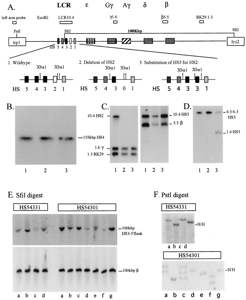
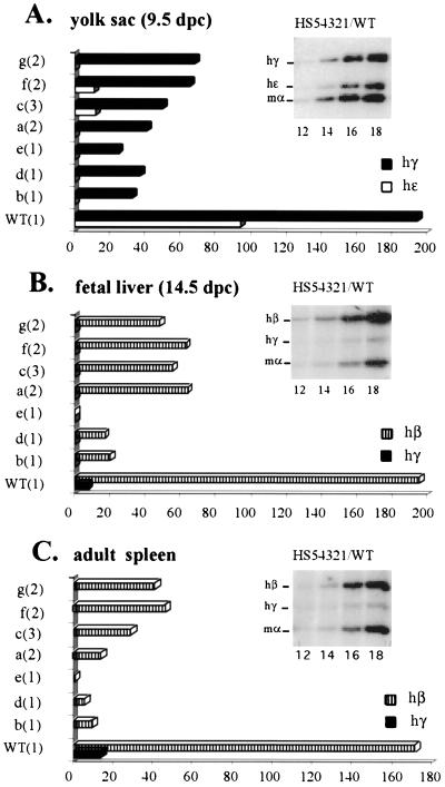
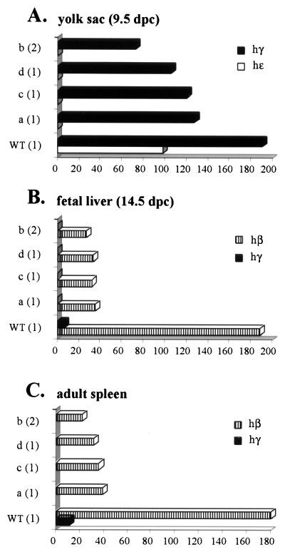
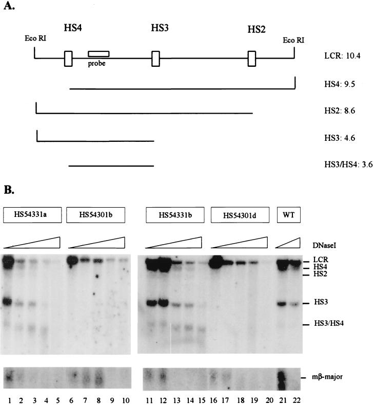
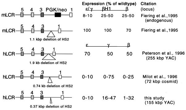
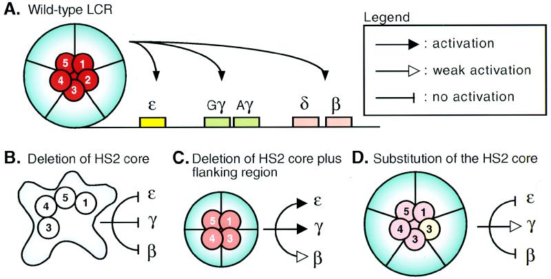
The human beta-globin locus control region (LCR) harbors both strong chromatin opening and enhancer activity when assayed in transgenic mice. To understand the contribution of individual DNase I hypersensitive sites (HS) to the function of the human beta-globin LCR, we have mutated the core elements within the context of a yeast artificial chromosome (YAC) carrying the entire locus and then analyzed the effect of these mutations on the formation of LCR HS elements and expression of the genes in transgenic mice. In the present study, we examined the consequences of two different HS2 mutations. We first generated seven YAC transgenic lines bearing a deletion of the 375-bp core enhancer of HS2. Single-copy HS2 deletion mutants exhibited severely depressed HS site formation and expression of all of the human beta-globin genes at every developmental stage, confirming that HS2 is a vital, integral component of the LCR. We also analyzed four transgenic lines in which the core element of HS2 was replaced by that of HS3 and found that while HS3 is able to restore the chromatin-opening activity of the LCR, it is not able to functionally replace HS2 in mediating high-level globin gene transcription. These results continue to support the hypothesis that HS2, HS3, and HS4 act as a single, integral unit to regulate human globin gene transcription as a holocomplex, but they can also be interpreted to say that formation of a DNase I hypersensitive holocomplex alone is not sufficient for mediating high-level globin gene transcription. We therefore propose that the core elements must productively interact with one another to generate a unique subdomain within the nucleoprotein holocomplex that interacts in a stage-specific manner with individual globin gene promoters.


Figures

FIG. 1
FIG. 1
Generation and structural analysis of HS2-mutant human β-globin YACs in yeast and transgenic mice. (A) Replacement of the 375-bp core enhancer of HS2 by the 220-bp core enhancer of HS3. Yeast cells were transformed with the targeting vectors (pRS306, linearized with AvaI, with or without HS3 embedded in HS2-flanking sequences). Yeast clones containing the vector integrated at the homologous site were grown in the presence of FOA, which selects for cells excising the URA3 marker together with either the mutant copy, thus restoring the wild-type configuration (1, HS54321), or together with the wild-type HS2 core element, generating a mutant LCR in which either HS2 is deleted (2, HS54301) or replaced by HS3 (3, HS54331), respectively. (B) Pulsed-field gel electrophoretic and Southern blot analysis of wild-type (lane 1) and mutant human β-globin YACs (HS54301 [lane 2] and HS54331 [lane 3]). Yeast chromosomes, embedded in agarose plugs, were loaded on a pulsed-field gel. After electrophoresis, the DNA was transferred to nylon membranes and hybridized to a radiolabeled 290-bp HS4 core enhancer fragment. (C and D) Southern blot analysis of yeast DNA carrying either wild-type (lane 1) or mutant YACs (HS54301 [lane 2] and HS54331 [lane 3]). Yeast DNA was isolated and digested with EcoRI (C) or XbaI (D). After electrophoresis the DNA was transferred to nylon membranes and hybridized with various radiolabeled probes derived from throughout the human β-globin gene locus (as indicated in panel A) (4). (E) Pulsed field gel electrophoretic and Southern blot analysis of the integrity of mutant human β-globin YAC transgenic mice (mutant lines HS54331a to HS54331d and HS54301a to HS54301g, as indicated). After electrophoresis, the DNA was transferred to nylon membranes and hybridized to radiolabeled probes derived from the 5′-flanking region of HS3 (top) or to a fragment corresponding to the second intron of the adult β-globin gene (bottom). (F) Analysis of the copy number of the mutant human β-globin YAC transgenes (same numbering as in panel E). Mouse genomic DNA, isolated from the tails of transgenic mice, was digested with PstI. After electrophoresis and transfer to nylon membranes, the DNA was hybridized to a 723-bp PstI-AwlI fragment derived from the left YAC vector arm (H/H indicates the presence of two left-arm fragments integrated into a head-to-head configuration in HS54331b, HS54301e, and HS54301g).
FIG. 2
FIG. 2
Analysis of globin gene expression in YAC transgenic mice bearing a deletion of the 375-bp core enhancer of HS2. RNA was extracted from yolk sac (at 9.5 d.p.c.), fetal liver (14.5 d.p.c.), or adult spleen (4 to 6 weeks) and subjected to RT-PCR analysis was performed with a mixture of globin primers (amplifying human ɛ-, γ-, and β-as well as mouse α-globin genes) and a radiolabeled nucleotide. After electrophoresis, samples (amplifying within the linear range) were analyzed and quantified by phosphorimaging, as described previously (4). The insert in each panel shows an autoradiograph depicting characteristic PCR products obtained from RNA extracted from the yolk sac (A), fetal liver (B), or anemic spleen (C) of transgenic embryos or mice carrying the wild-type β-globin YAC HS54321 (cycle numbers are indicated below the lanes). (A) Expression profile of human globin genes in the embryonic yolk sac taken at 9.5 d.p.c. (B) Expression profile of human globin genes in the fetal liver taken at 14.5 d.p.c. (C) Expression profile of human β-globin genes in the adult spleen taken at from 4- to 6-week-old anemic mice. The lines analyzed in each columns are indicated on the left of each panel with the transgene copy numbers shown in parentheses (WT, wild-type HS54321; a to g, HS54301 mutant lines). Levels of individual globin gene transcription are expressed as a percentage of mouse α-globin gene expression (which was set at 100%) per gene copy.
FIG. 3
FIG. 3
Analysis of YAC transgenic mice in which the 375-bp core enhancer of HS2 is replaced by the 220-bp core element of HS3. RNA was analyzed at the various stages of mouse hematopoiesis as described in the legend to Fig. 2. Shown are the expression profiles of the human globin genes in the embryonic yolk sac (9.5 d.p.c.) (A), fetal liver (14.5 d.p.c.) (B), and adult spleen (C). Indicated on the left of each panel is the specific line analyzed in each column (WT, transgene carrying wild-type YAC HS54331; a to d, HS54331 mutant lines). Numbers in parentheses are the copy numbers of the YACs transgenes. Expression is based on levels of mouse α-globin (set at 100%) calculated per integrated gene copy.
FIG. 4
FIG. 4
DNase I HS mapping of the human β-globin LCR in HS2 mutant and wild-type human β-globin YAC transgenic mice. Nuclei were isolated from spleens of anemic transgenic mice bearing wild-type or HS2 mutant human β-globin YACs. The nuclei were incubated with increasing concentrations of DNase I, and the DNA was isolated and digested with EcoRI. After gel electrophoresis the DNA was hybridized to radioactively labeled probes corresponding to the 3′-flanking region of HS4 (as indicated in panel A) or to the 5′ promoter region of the mouse βmajor-globin gene. (A) Diagrammatic representation of the fragments generated by DNase I and EcoRI in the human β-globin LCR (indicated on the right are the sizes, in kilobase pairs of the various fragments). (B) HS formation in the wild-type (WT) or HS2 mutant human β-globin YAC transgenic mice. Two lines representing each mutant were analyzed for HS formation within the LCR, the substitution lines HS54331a and HS54331b (lanes 1 to 5 and 11 to 15, respectively) and two deletion mutants HS54301b and HS54301d (lanes 6 to 10 and 16 to 20, respectively). Lanes 21 and 22 show hypersensitivity in the single-copy transgene carrying the wild-type human β-globin YAC (HS54321a) (4). As a control for the DNase I digest, the nylon membranes were rehybridized to a probe detecting a characteristic HS site in the mouse βmajor-globin gene promoter (shown at the bottom of the picture).
FIG. 5
FIG. 5
Comparison of results obtained from studies examining deletion of HS2 from the endogenous mouse or from the human β-globin gene loci. Expression levels of individual globin genes were calculated as percentages of expression levels of particular genes expressed from the wild-type loci (set at 100%).
FIG. 6
FIG. 6
HS core elements form a synergistic active subdomain within the LCR holocomplex. The figure depicts a cartoon of a model consistent with our data. (A) The LCR in the wild-type configuration. The core elements, together with the flanking regions, generate the holocomplex (light blue), with the core elements folded together to establish the specific configuration required for the active site (red). (B) Deletion of the HS2 core enhancer results in collapse of the LCR holocomplex and active site and renders the LCR unable to confer high-level or transgene integration position-independent transcription to the globin genes. (C) Deletion of the HS2 core element together with its flanking sequences allows the formation of an imperfect, alternative structure for the holocomplex, which retains transgene integration site-independent transcription but is impaired in its activity provided by the subdomain (light red). (D) Replacement of the HS2 by the HS3 core element allows the formation of an essentially wild-type holocomplex (thus retaining position-independent transcription of the integrated transgenes), but the active site is again unable to stimulate all the different globin genes at each developmental stage because it (light red) is not perfectly re-formed at each stage.

References

    1. Aladjem M I, Groudine M, Brody L L, Dieken E S, Fournier R E, Wahl G M, Epner E M. Participation of the human β-globin locus control region in initiation of DNA replication. Science. 1995;270:815–819. - PubMed
    1. Bonifer C, Vidal M, Grosveld F, Sippel A E. Tissue specific and position independent expression of the complete gene domain for chicken lysozyme in transgenic mice. EMBO J. 1990;9:2843–2848. - PMC - PubMed
    1. Bresnick E, Tze L. Synergism between hypersensitive sites confers long-range gene activation by the β-globin locus control region. Proc Natl Acad Sci USA. 1997;94:4566–4571. - PMC - PubMed
    1. Bungert J, Dave U, Lieuw K E, Lim K-C, Shavit J A, Liu Q, Engel J D. Synergistic regulation of human β-globin gene switching by locus control region elements HS3 and HS4. Genes Dev. 1995;9:3083–3096. - PubMed
    1. Caterina J, Ryan T M, Pawlik R D, Brinster R L, Behringer R R, Townes T M. Human β-globin locus control region: analysis of the 5′ DNase I hypersensitive site HS 2 in transgenic mice. Proc Natl Acad Sci USA. 1991;88:1626–1630. - PMC - PubMed

Publication types

MeSH terms

LinkOut - more resources