. 1999 Apr;19(4):3103-14.
doi: 10.1128/MCB.19.4.3103.

Control of replicative life span in human cells: barriers to clonal expansion intermediate between M1 senescence and M2 crisis

Affiliations

Control of replicative life span in human cells: barriers to clonal expansion intermediate between M1 senescence and M2 crisis

J A Bond et al. Mol Cell Biol. 1999 Apr.

Abstract

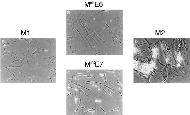
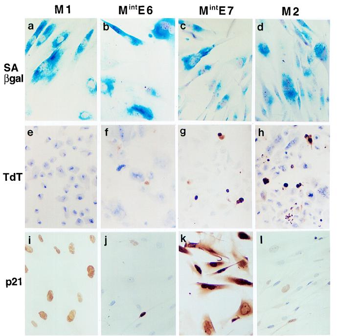
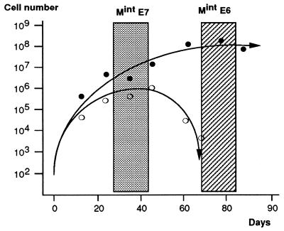
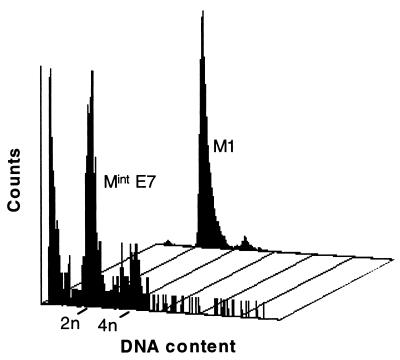
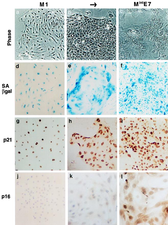
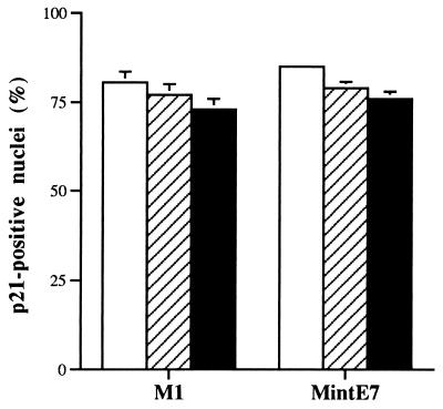
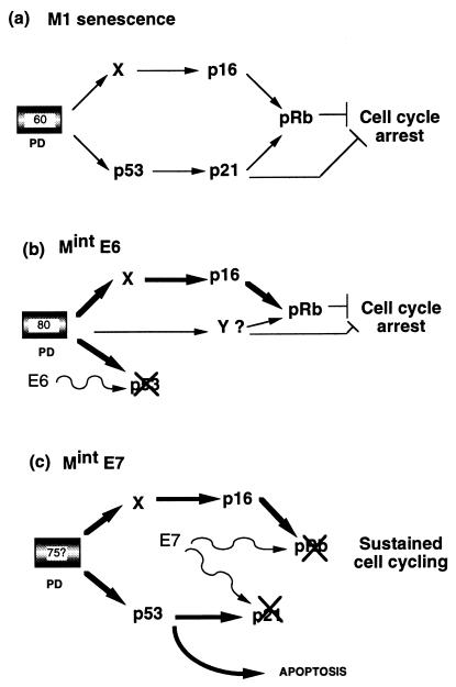
The accumulation of genetic abnormalities in a developing tumor is driven, at least in part, by the need to overcome inherent restraints on the replicative life span of human cells, two of which-senescence (M1) and crisis (M2)-have been well characterized. Here we describe additional barriers to clonal expansion (Mint) intermediate between M1 and M2, revealed by abrogation of tumor-suppressor gene (TSG) pathways by individual human papillomavirus type 16 (HPV16) proteins. In human fibroblasts, abrogation of p53 function by HPVE6 allowed escape from M1, followed up to 20 population doublings (PD) later by a second viable proliferation arrest state, MintE6, closely resembling M1. This occurred despite abrogation of p21(WAF1) induction but was associated with and potentially mediated by a further approximately 3-fold increase in p16(INK4a) expression compared to its level at M1. Expression of HPVE7, which targets pRb (and p21(WAF1)), also permitted clonal expansion, but this was limited predominantly by increasing cell death, resulting in a MintE7 phenotype similar to M2 but occurring after fewer PD. This was associated with, and at least partly due to, an increase in nuclear p53 content and activity, not seen in younger cells expressing E7. In a different cell type, thyroid epithelium, E7 also allowed clonal expansion terminating in a similar state to MintE7 in fibroblasts. In contrast, however, there was no evidence for a p53-regulated pathway; E6 was without effect, and the increases in p21(WAF1) expression at M1 and MintE7 were p53 independent. These data provide a model for clonal evolution by successive TSG inactivation and suggest that cell type diversity in life span regulation may determine the pattern of gene mutation in the corresponding tumors.


Figures

FIG. 1
FIG. 1
Comparison of Mint phenotypes with M1 (senescence) and M2 (crisis) in HDF. Near-senescent cultures of HDF were infected with retroviral vectors expressing neo only (control) (A), HPVE6 (B), HPVE7 (C), or HPVE6 plus E7 (D) and passaged until they entered a state of zero net growth, designated respectively M1, MintE6, MintE7, and M2. Phase-contrast photomicrographs of MintE6 cultures (B) show cell flattening with very few mitoses or dying cells, a phenotype closely resembling M1 (A). In contrast, MintE7 (C) shows continuing cell turnover resembling M2 (D). Magnification, ×100.
FIG. 2
FIG. 2
Further characterization of Mint phenotypes in human fibroblasts. (a to d) Histochemical assessment of SAβ-gal activity showing very high expression in M1 (a), MintE6 (b), MintE7 (c), and M2 (d) growth arrest states. (e to h) TdT assay showing frequent apoptosis in MintE7 (g) and M2 (h) states but not at MintE6 (f) or M1 (e). (i to l) Immunocytochemical analysis of p21WAF1 protein showing further elevation of expression at MintE7 (k) above that at M1 (i) contrasting with reduced levels at MintE6 (j) and M2 (l). Hematoxylin counterstain; magnification, ×100.
FIG. 3
FIG. 3
Contrasting kinetics of growth arrest at MintE6 and MintE7. Approximately 2 × 10⁵ HDF at <3 PD from senescence were infected with vectors expressing neo plus either HPV E6 (solid symbols) or E7 (open symbols), selected in G418 and maintained in culture with passage as necessary for up to 3 months. Changes in total population size were calculated from passage ratios, and cell counts were performed at the indicated intervals. The initial number of stably infected G418-resistant cells at time 0 was estimated from the colony yield in parallel experiments conducted at clonal density. (No direct cell counts were possible until G418 selection was complete by day 10.) Both E6- and E7-expressing cultures escape senescence and grow for several weeks. With E6, this is followed by a stable state of growth arrest, referred to here as MintE6 (hatched zone). In contrast, E7 cultures show only a temporary stationary-growth phase, termed here MintE7 (shaded zone), which is followed by progressive net cell loss. These boxed zones of the growth curves define the phases at which cultures were used for all other analyses of the Mint phenotypes. Results shown are means of two representative experiments in each case, together with best-fit curves.
FIG. 4
FIG. 4
Flow cytometric analysis of DNA content in human fibroblasts at M1 and MintE7. Note prominent sub-G1 peak in MintE7, indicative of apoptosis.
FIG. 5
FIG. 5
Western blot analysis of p21WAF1 and p16INK4a proteins in fibroblast populations expressing HPV16E6 and/or E7. (a) p21 protein was expressed at higher levels in MintE7 cultures (lane 5) and at lower levels in MintE6 (lane 4) and M2 (lane 6) cultures compared to a control (neo) M1 population (lane 3). Young cycling fibroblasts at PDL ∼25 (lane 1) and uninfected fibroblasts at M1 (lane 2) are shown for comparison. (b) p16 protein was expressed at higher levels in all Mint and M2 cultures than at M1. Contents of lanes are the same as those in panel a. Equal amounts (30 μg) of lysate protein were loaded per lane, as verified by India ink staining shown below each blot. Molecular mass markers are shown in kilodaltons.
FIG. 6
FIG. 6
Increased expression of p53 protein in post-M1 fibroblasts and thyroid epithelial cells expressing HPVE7. Immunocytochemical analysis with PAb421 antibody fails to detect p53 in most young HDF (PDL, ∼25), both in neo-only controls (c) and in cells expressing HPVE7 (d). In contrast, readily detectable nuclear p53 with a characteristic cell-cell heterogeneity is seen in HDF expressing E7 at MintE7 (f) but not in neo-only cells at M1 (e). These differences in p53 content are not explicable by differences in E7 protein levels, which are similar in both young (a) and post-M1 (b) HDF expressing the E7 vector. A similar heterogeneous elevation of p53 levels is seen in human thyrocytes whose life span has been extended by expression of E7 (h) but not in neo controls at the end of their normal proliferative life span (g). Immunoperoxidase with hematoxylin counterstain; magnification, ×125.
FIG. 7
FIG. 7
Phenotypes of human thyroid epithelial cells expressing HPVE7. (a to j) Normal untreated cultures at the end of their normal proliferative life span (M1); (b to k) proliferating colonies induced by expression of HPVE7; (c to l) E7-expressing cultures after net growth has ceased (MintE7). All stages show high levels of SAβ-gal (d to f). p21WAF1 is readily detectable by immunocytochemistry in most nuclei at M1 (g) and is further induced following extension of life span by E7, even while colonies are still proliferating (h). p16 is undetectable at M1 and in E7-expressing cells is induced later than p21, (l). Note that although terminal growth arrest in normal thyrocytes, or M1, occurs after just a few PD, it may not be triggered by the same underlying regulator as that in HDF. Phase-contrast micrographs (a to c); histochemical β-Gal assay (d to f); immunoperoxidase with hematoxylin counterstain (g to l). Magnification, ×90.
FIG. 8
FIG. 8
p53-independent expression of p21waf-1 in both normal and HPVE7-expressing thyroid epithelial cells. Thyrocytes at the end of either their normal proliferative lifespan (M1) or the extended life span conferred by HPVE7 (MintE7) were microinjected with control IgG (open bars), anti-p53 antibodies PAb1801 (hatched bars), or DO-1 (solid bars). p21 was analyzed 72 h later by immunofluorescence, and the percentage of immunopositive nuclei was counted. Results are shown as means of three replicate experiments ± standard errors of the mean.
FIG. 9
FIG. 9
Models to explain Mint growth arrest states in human fibroblasts. (a) Normal senescence (M1) is assumed to be mediated by at least two signal pathways activated by a cell-cycle clock after around 60 PD (now almost certainly linked to telomere erosion). Both of these converge to inhibit phosphorylation of Rb by CDKs. p21 may also inhibit cell cycle progression via other targets (51). (b) MintE6. Cells which have initially escaped senescence by E6-mediated loss of the p53 pathway are able to reestablish cell cycle arrest approximately 20 PD later. One mechanism is further upregulation of the p16 pathway, which may compensate for the loss of p21, although our data do not exclude the requirement for an additional, as-yet-unknown, pathway(s) indicated by Y. (c) MintE7. Escape from senescence induced by HPVE7 is associated with a more profound disruption of cell cycle control through abrogation of pRb and p21 (22, 30a), which prevents restoration of cell cycle arrest. Clonal expansion is finally limited by apoptosis mediated at least in part by p53.

References

    1. Afshari C A, Vojta P J, Annab L A, Futreal P A, Willard T B, Barrett J C. Investigation of the role of G1/S cell cycle mediators in cellular senescence. Exp Cell Res. 1993;209:231–237. - PubMed
    1. Alcorta D A, Xiong Y, Phelps D, Hannon G, Beach D, Barrett J C. Involvement of the cyclin-dependent kinase inhibitor p16 (INK4a) in replicative senescence of normal human fibroblasts. Proc Natl Acad Sci USA. 1996;93:13742–13747. - PMC - PubMed
    1. Atadja P, Wong H, Garkavtsev I, Geillette C, Riabowol K. Increased activity of p53 in senescing fibroblasts. Proc Natl Acad Sci USA. 1995;92:8348–8352. - PMC - PubMed
    1. Bacchetti S. Telomere dynamics and telomerase activity in cell senescence and cancer. Cell Dev Biol. 1996;7:31–39.
    1. Banks L, Matlashewski G, Crawford L. Isolation of human p53 specific monoclonal antibodies and their use in the study of human p53 expression. Eur J Biochem. 1986;159:529–534. - PubMed

Publication types

MeSH terms

Substances