. 1999 Apr;67(4):1757-62.
doi: 10.1128/IAI.67.4.1757-1762.1999.

A novel urease-negative Helicobacter species associated with colitis and typhlitis in IL-10-deficient mice

Affiliations

A novel urease-negative Helicobacter species associated with colitis and typhlitis in IL-10-deficient mice

J G Fox et al. Infect Immun. 1999 Apr.

Abstract

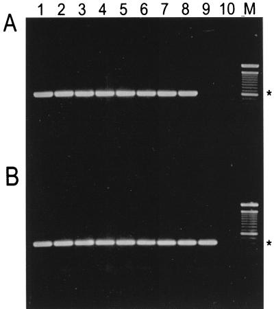
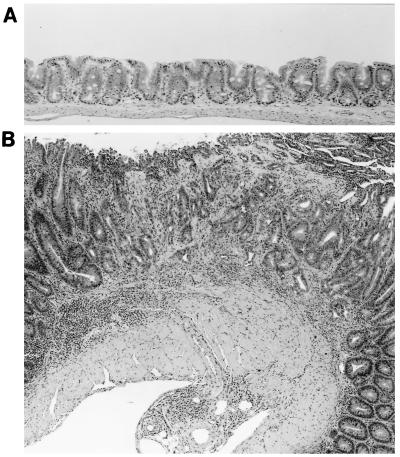
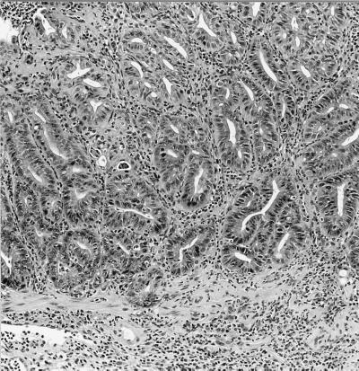
A spiral-shaped bacterium with bipolar, single-sheathed flagella was isolated from the intestines of IL-10 (interleukin-10)-deficient (IL-10(-/-)) mice with inflammatory bowel disease. The organism was microaerobic, grew at 37 and 42 degrees C, and was oxidase and catalase positive but urease negative. On the basis of 16S rRNA gene sequence analysis and biochemical and phenotypic criteria, the organism is classified as a novel helicobacter. Cesarean section-rederived IL-10(-/-) mice without helicobacter infection did not have histological evidence of intestinal inflammation. However, helicobacter-free IL-10(-/-), SCID/NCr, and A/JNCr mice experimentally inoculated with the novel urease-negative Helicobacter sp. developed variable degrees of inflammation in the lower intestine, and in immunocompetent mice, the experimental infection was accompanied by a corresponding elevated immunoglobulin G antibody response to the novel Helicobacter sp. antigen. These data support other recent studies which demonstrate that multiple Helicobacter spp. in both naturally and experimentally infected mice can induce inflammatory bowel disease. The mouse model of helicobacter-associated intestinal inflammation should prove valuable in understanding how specific microbial antigens influence a complex disease process.


Figures

FIG. 1
FIG. 1
Phylogenetic tree constructed on the basis of 16S rRNA sequence similarity values, using the neighbor-joining method. Scale bar = 5% difference in nucleotide sequences as determined by measuring the lengths of the horizontal lines connecting two species.
FIG. 2
FIG. 2
Phosphotungstic acid negative stain of urease-negative Helicobacter sp. depicting helical bacteria with bipolar sheathed flagellae. Magnification, ×13,650.
FIG. 3
FIG. 3
Agarose gel electrophoresis of DNA amplified by PCR with Helicobacter species-specific primers JGF-F1 and JGF-R1 (A) or Helicobacter genus-specific primers C97 and C98 (B). Lanes 1 to 8, novel urease-negative Helicobacter sp. isolates MIT 97-6810 and MIT 97-6811 and isolates 98-6781, 98-6782, 98-784, 98-9785, 98-7686, and 98-6787, respectively; lane 9, H. rodentium 95-1707; lane 10, control with no DNA template; M, 100-bp DNA ladder (GibcoLife Technologies, Gaithersburg, Md.). The 0.6-kb (A) and 0.4-kb (B) positions are indicated by asterisks.
FIG. 4
FIG. 4
U HelAg-specific Ab in sera of urease-negative Helicobacter-infected mice. Sera from 5.5-month-infected A/J (●) and SCID (▴) mice (A) and from 4.5-month-infected IL-10−/− (■) mice (B) were analyzed for levels of total IgG reactive to U HelAg by ELISA as described in Materials and Methods. Symbols represent means ± standard deviations of duplicate ELISA values from pools of three infected A/J, two infected SCID, and seven infected IL-10−/− mice, respectively. Uninfected IL-10−/− mice showed no reactivity to the U HelAg (data from a separate experiment, not shown). OD, optical density.
FIG. 5
FIG. 5
Diffuse and focal epithelial hyperplasia with marked inflammation in the cecum of a IL-10−/− mouse naturally infected with the urease-negative Helicobacter sp. H&E; magnification, ×30.
FIG. 6
FIG. 6
(A) Normal cecum of a 4-month-old uninfected control IL-10−/− mouse. H&E stain × 75. (B) Focal atypical hyperplasia and diffuse hyperplasia with marked chronic inflammation in cecum of an IL-10−/− mouse, 6 months after infection with the novel urease-negative Helicobacter sp. H&E; magnification, ×30.
FIG. 7
FIG. 7
Focal atypical hyperplasia and chronic inflammation in rectum of an IL-10−/− mouse, 6 months after injection with the novel urease-negative Helicobacter sp. H&E; magnification, ×75.

References

    1. Berg O J, Davidson N, Kuhn R, Muller W, Menon S, Holland G, Thompson-Snipes L. Enterocolitis and colon cancer in interleukin 10 deficient mice are associated with aberrant cytokine production and CD4 + Th1-like response. J Clin Investig. 1996;98:1010–1020. - PMC - PubMed
    1. Cahill R J, Foltz C J, Fox J G, Dangler C A, Powrie F, Schauer D B. Inflammatory bowel disease: an immune-mediated condition triggered by bacterial infection with Helicobacter hepaticus. Infect Immun. 1997;65:3126–3131. - PMC - PubMed
    1. Chin E Y, Jha J, Dangler C A, Schauer D B. Abstracts of the 98th General Meeting of the American Society for Microbiology 1998. Washington, D.C: American Society for Microbiology; 1998. Infectious colitis in T cell receptor knockout mice, abstr. B436.
    1. Foltz C J, Fox J G, Cahill R J, Murphy R C, Yan L, Shames B, Schauer D B. Spontaneous inflammatory bowel disease in multiple mutant mouse lines: association with colonization by Helicobacter hepaticus. Helicobacter. 1998;3:69–78. - PubMed
    1. Fox J G, Dewhirst F E, Tully J G, Paster B J, Yan L, Taylor N S, Collins M J, Gorelick P L, Ward J M. Helicobacter hepaticus sp. nov., a microaerophilic bacterium isolated from livers and intestinal mucosal scrapings from mice. J Clin Microbiol. 1994;32:1238–1245. - PMC - PubMed

Publication types

MeSH terms

Associated data

LinkOut - more resources