. 1999 Apr;67(4):1779-88.
doi: 10.1128/IAI.67.4.1779-1788.1999.

Extracellular cysteine protease produced by Streptococcus pyogenes participates in the pathogenesis of invasive skin infection and dissemination in mice

Affiliations

Extracellular cysteine protease produced by Streptococcus pyogenes participates in the pathogenesis of invasive skin infection and dissemination in mice

S Lukomski et al. Infect Immun. 1999 Apr.

Abstract

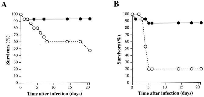
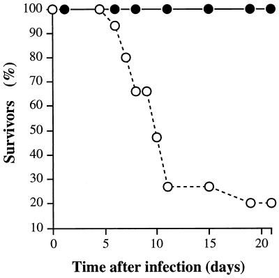
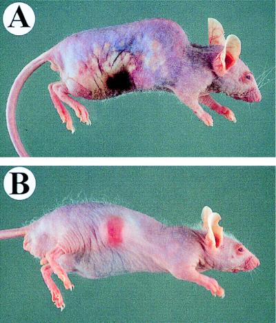
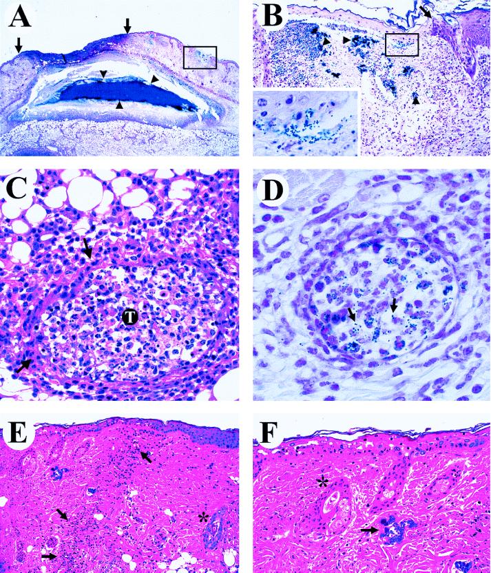
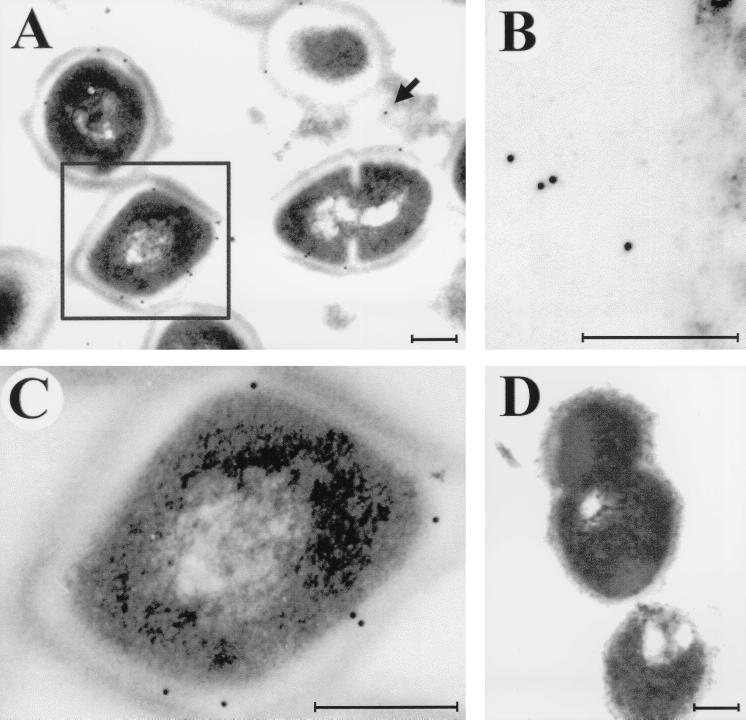
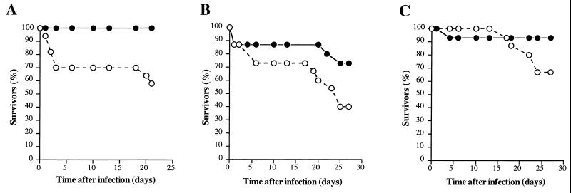
The role of an extracellular cysteine protease encoded by the speB gene in group A Streptococcus (GAS) skin infection was studied with a mouse model. Mice were injected subcutaneously with a wild-type GAS serotype M3 strain or a cysteine protease-inactivated isogenic derivative grown to stationary phase. The mortality rate of mice injected with the M3 speB mutant strain was significantly decreased (P < 0.0008) compared to that of animals injected with the wild-type parental organism. The abscesses formed in animals infected with the cysteine protease mutant strain were significantly smaller (P < 0.0001) than those caused by the wild-type organism and slowly regressed over 3 to 4 weeks. In striking contrast, infection with the wild-type GAS isolate generated necrotic lesions, and in some animals the GAS disseminated widely from the injection site and produced extensive cutaneous damage. All of these animals developed bacteremia and died. GAS dissemination was accompanied by severe tissue and blood vessel necrosis. Cysteine protease expression in the infected tissue was identified by immunogold electron microscopy. These data demonstrate that cysteine protease expression contributes to soft tissue pathology, including necrosis, and is required for efficient systemic dissemination of the organism from the initial site of skin inoculation.


Figures

FIG. 1
FIG. 1
Kaplan-Meier survival curves (n = 15 mice in each group) following subcutaneous inoculation with the wild-type Streptococcus pyogenes serotype M3 strain (open circles) and the cysteine protease-inactivated isogenic M3 speB derivative (solid circles) grown overnight. (A) Inoculum of ∼107 CFU; χ2 = 4.2 and P < 0.0398. (B) Inoculum of ∼108 CFU; χ2 = 11.2 and P < 0.0008.
FIG. 2
FIG. 2
Kaplan-Meier survival curves (n = 15 mice in each group) following intranasal inoculation with the wild-type S. pyogenes serotype M3 strain (open circles) and the cysteine protease-inactivated isogenic M3 speB derivative (solid circles). A significant difference in mouse mortality was observed (inoculum, ∼107 CFU; χ2 = 20.2 and P < 0.0001).
FIG. 3
FIG. 3
Cutaneous lesions in mice inoculated with wild-type and isogenic speB mutant GAS strains. (A) Mouse infected subcutaneously with 107 CFU of the wild-type M3 isolate. The infection spread radially (day 3) from the inoculation site and resulted in extensive subcutaneous and dermal necrosis involving a large portion of the lateral side of the animal. (B) Mouse inoculated with 107 CFU of the cysteine protease-inactivated M3 mutant strain. A solitary subcutaneous abscess formed and then regressed over time.
FIG. 4
FIG. 4
Photomicrographs demonstrating different aspects of the skin pathology in animals infected with the wild-type strain expressing cysteine protease. (A and B) Bacterial spread from the initial site of injection (Gram stain). (A) A large depressed cutaneous infarct is located between the solid arrows. Note the large colony of gram-positive (blue) cocci in the center of a solitary subcutaneous abscess (arrowheads). One of several microinfarcts is boxed. Magnification, ×4. (B) Photomicrograph of one microinfarct at a higher magnification (×41). The arrow identifies the junction of an epidermal infarct, with healthy tissue located on the right and a pale pink necrotizing epidermitis on the left. Note blue-stained bacterial colonies in the dermis (arrowheads). The inset shows a higher magnification (×164) of the boxed area and demonstrates the spread of gram-positive cocci into the upper dermis and epidermis. (C and D) Bacterial spreading results in vascular pathology. (C) Necrotizing vasculitis of a subcutaneous blood vessel (arrows) with a thrombus (T) (hematoxylin and eosin stain). The vascular lumen contains fibrin, neutrophils, and necrotic cellular debris. Magnification, ×82. (D) A blood vessel with early thrombosis and numerous gram-positive-cocci (arrows) located within the thrombus (Gram stain). Magnification, ×164. (E and F) Bacterial spreading causes cutaneous infarction (hematoxylin and eosin stain). (E) A line of demarcation (arrows) is located between healthy skin on the right and the necrotic zone on the left, with linear infiltration of polymorphonuclear leukocytes at the interface. Note the normal blue-stained hair follicle within a histologically normal zone (*). Magnification, ×41. (F) Higher magnification (×82) demonstrating necrosis of basal cells and pallor of the stratum granulosum and stratum spinosum in the epidermal infarct. Note blue-stained bacterial colonies in the upper dermis (arrow) and the abnormal light pink staining of the hair follicle in the infarcted area (*).
FIG. 5
FIG. 5
Immunogold electron microscopy localization of the cysteine protease produced by the wild-type GAS during skin infection. (A) Cysteine protease is produced by GAS within infected tissue; the cysteine protease fraction is detected in a secreted form (solid arrow) in the tissue or as a fraction still associated with GAS cells (boxed cell). (B) Cysteine protease fraction released into surrounding tissue. (C) Cell-associated cysteine protease fraction of the boxed cell in panel A. (D) Negative control in which no nonspecific background labeling is detected. Tissue samples were collected on day 2 following inoculation with 108 CFU of the wild-type M3 strain. Bars, 200 nm.
FIG. 6
FIG. 6
Kaplan-Meier survival curves (n = 15 mice in each group) following subcutaneous inoculation with the wild-type S. pyogenes serotype M3 strain (open circles) and the cysteine protease-inactivated isogenic M3 speB derivative (solid circles). (A) Inoculum prepared from early-log-phase cultures (OD600 ∼ 0.25); 1.3 × 10⁸ CFU of the wild-type M3 and 2.1 × 10⁸ CFU of the M3 speB mutant were injected (χ2 = 7.6 and P < 0.0005). (B) Inoculum prepared from middle-log-phase cultures (OD600 ∼ 0.4); 1.9 × 10⁸ CFU of the wild-type M3 and 2.5 × 10⁸ CFU of the M3 speB mutant were injected (χ2 = 2.9 and P < 0.0834). (C) Inoculum prepared from very-late-phase cultures (OD600 ∼ 0.8); 1.8 × 10⁸ CFU of the wild-type M3 and 1.8 × 10⁸ CFU of the M3 speB mutant were injected (χ2 = 2.9 and P < 0.0859).

References

    1. Ashbaugh C D, Warren H B, Carey V J, Wessels M R. Molecular analysis of the role of the group A streptococcal cysteine protease, hyaluronic acid capsule, and M protein in a murine model of human invasive soft-tissue infection. J Clin Investig. 1998;102:550–560. - PMC - PubMed
    1. Barker F G, Leppard B J, Seal D V. Streptococcal necrotising fasciitis: comparison between histological and clinical features. J Clin Pathol. 1987;40:335–341. - PMC - PubMed
    1. Belani K, Schlievert P M, Kaplan E L, Ferrieri P. Association of exotoxin-producing group A streptococci and severe disease in children. Pediatr Infect Dis J. 1991;10:351–354. - PubMed
    1. Boyle M D P, Raeder R, Flosdorff A, Podbielski A. Role of emm and mrp genes in the virulence of group A streptococcal isolate 64/14 in a mouse model of skin infection. J Infect Dis. 1998;177:991–997. - PubMed
    1. Burns E H, Jr, Lukomski S, Rurangirwa J, Podbielski A, Musser J M. Genetic inactivation of the extracellular cysteine protease enhances in vitro internalization of group A streptococci by human epithelial and endothelial cells. Microb Pathog. 1998;24:333–339. - PubMed

Publication types