. 1999 Apr;67(4):1910-6.
doi: 10.1128/IAI.67.4.1910-1916.1999.

Characterization of a novel trypanosome lytic factor from human serum

Affiliations

Characterization of a novel trypanosome lytic factor from human serum

J Raper et al. Infect Immun. 1999 Apr.

Abstract

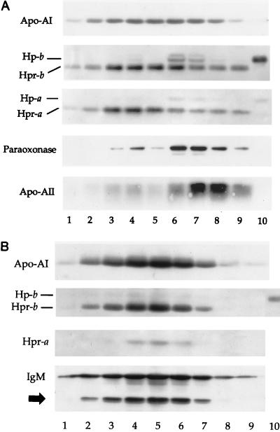
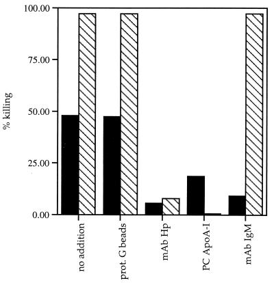
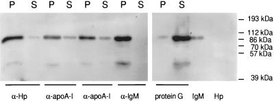
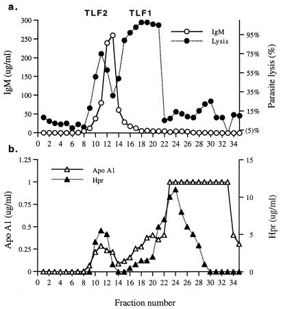
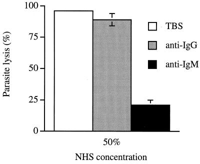
Natural resistance of humans to the cattle pathogen Trypanosoma brucei brucei has been attributed to the presence in human serum of nonimmune factors that lyse the parasite. Normal human serum contains two trypanosome lytic factors (TLFs). TLF1 is a 500-kDa lipoprotein, which is reported to contain apolipoprotein A-I (apoA-I), haptoglobin-related protein (Hpr), hemoglobin, paraoxonase, and apoA-II, whereas TLF2 is a larger, poorly characterized particle. We report here a new immunoaffinity-based purification procedure for TLF2 and TLF1, as well as further characterization of the components of each purified TLF. Immunoaffinity-purified TLF1 has a specific activity 10-fold higher than that of TLF1 purified by previously described methods. Moreover, we find that TLF1 is a lipoprotein particle that contains mainly apoA-I and Hpr, trace amounts of paraoxonase, apoA-II, and haptoglobin, but no detectable hemoglobin. Characterization of TLF2 reveals that it is a 1,000-kDa protein complex containing mainly immunoglobulin M, apoA-I, and Hpr but less than 1% detectable lipid.


Figures

FIG. 1
FIG. 1
Silver-stained SDS–12% polyacrylamide gel of reduced TLF1 and TLF2. One lytic unit of TLF2 and 7.5 lytic units of TLF1 were reduced in sample buffer, and the polypeptides were separated by PAGE. Bands identified by protein sequencing of their N termini (heavy chain [H-chain] and light chain [L-chain]) are indicated with white arrows. We could not obtain the sequences of the two bands indicated with the black arrow but they may be proteolytic products of the IgM μ chain due to their immunoreactivity with anti-μ-chain polyclonal antibody (see Fig. 2). Asterisks indicate positions of molecular weight markers migrating at 94, 67, 43, 30, 20.1, and 14 kDa (top to bottom).
FIG. 2
FIG. 2
Western blot of fractions from final step of purification of TLF1 (A) and TLF2 (B). Superose 6HR fractions encompassing molecular mass ranges of 650 to 200 kDa for TLF1 (A, lanes 1 to 9) and 1,200 to 700 kDa for TLF2 (B, lanes 1 to 9) were reduced in sample buffer. The polypeptides were separated by SDS-PAGE prior to transfer to PVDF membranes. The blots were sequentially immunoblotted with antibodies raised against apoA-I, apoA-II, Hp and Hpr, paraoxonase, and IgM as indicated on LHS. The blots were stripped between each immunoblotting step by incubation in 0.2 M glycine (pH 2.8). The black arrow corresponds to that in Fig. 1. Lanes 10, haptoglobin standard.
FIG. 3
FIG. 3
Immunodepletion of TLF activity. Protein G beads (50 μl) were preincubated with 100 μg of the various antibodies. Following washing, 1 to 2 lytic units of TLF was added to the beads and incubated for 60 min at 4°C. The supernatants were then tested for trypanolytic activity. Shown are lytic activities of TLF2 (solid bars) and TLF1 (hatched bars) remaining after no addition (control) and after immunoprecipitation with protein (prot.) G beads only, anti-Hp MAb, goat anti-apoA-I polyclonal (PC) antibody, and anti-IgM MAb.
FIG. 4
FIG. 4
TLF2 is a protein complex. One microgram of TLF2 was immunoprecipitated by antibodies to human Hp (MAb; α-Hp), human apoA-I (polyclonal antibodies from sheep and goat; α-apoA-I), and human IgM (MAb; α-IgM). Following immunoprecipitation, the pellet (P) and supernatant (S) were separated by reducing SDS-PAGE (12% gel) and transferred to PVDF membranes. The IgM μ chain was detected with rabbit anti-IgM μ-chain antibody followed by goat anti-rabbit IgG-HRP and exposed for 30 s by ECL.
FIG. 5
FIG. 5
Analysis of TLF2 component proteins in serum fractions. NHS was fractionated by size exclusion chromatography, and collected fractions were assayed for trypanolytic activity (a) and indicated proteins (a and b).
FIG. 6
FIG. 6
Trypanolytic activity of IgM- and IgG-depleted NHS. Error bars show means ± standard deviation (n = 3).

References

    1. Aaronovitch S, Terry R J. The trypanolytic factor in human serum. Trans R Soc Trop Med Hyg. 1972;66:344. - PubMed
    1. Aszalos B F, Sloop C H, Wong L, Roheim P S. Two-dimensional electrophoresis of plasma lipoproteins: recognition of new apo A-I-containing subpopulations. Biochim Biophys Acta. 1993;1169:291–300. - PubMed
    1. Barth P. A new method for the isolation of the trypanocidal factor from normal human serum. Acta Trop. 1989;46:71–73. - PubMed
    1. Bensi G, Raugei G, Klefenz H, Cortese R. Structure and expression of the human haptoglobin locus. EMBO J. 1985;4:119–126. - PMC - PubMed
    1. Bowman B H. Hepatic plasma proteins: mechanism of function and regulation. San Diego, Calif: Academic Press, Inc.; 1993. pp. 159–167.

Publication types

LinkOut - more resources