. 1999 Mar 8;144(5):869-81.
doi: 10.1083/jcb.144.5.869.

SNARE membrane trafficking dynamics in vivo

Affiliations

SNARE membrane trafficking dynamics in vivo

D S Chao et al. J Cell Biol. .

Abstract

The ER/Golgi soluble NSF attachment protein receptor (SNARE) membrin, rsec22b, and rbet1 are enriched in approximately 1-micrometer cytoplasmic structures that lie very close to the ER. These appear to be ER exit sites since secretory cargo concentrates in and exits from these structures. rsec22b and rbet1 fused to fluorescent proteins are enriched at approximately 1-micrometer ER exit sites that remained more or less stationary, but periodically emitted streaks of fluorescence that traveled generally in the direction of the Golgi complex. These exit sites were reused and subsequent tubules or streams of vesicles followed similar trajectories. Fluorescent membrin- enriched approximately 1-micrometer peripheral structures were more mobile and appeared to translocate through the cytoplasm back and forth, between the periphery and the Golgi area. These mobile structures could serve to collect secretory cargo by fusing with ER-derived vesicles and ferrying the cargo to the Golgi. The post-Golgi SNAREs, syntaxin 6 and syntaxin 13, when fused to fluorescent proteins each displayed characteristic patterns of movement. However, syntaxin 13 was the only SNARE whose life cycle appeared to involve interactions with the plasma membrane. These studies reveal the in vivo spatiotemporal dynamics of SNARE proteins and provide new insight into their roles in membrane trafficking.


Figures

Figure 1
Figure 1
Schematic of reagents used in this study. mAbs and pAbs against the cytoplasmic domains of several SNARE proteins were characterized previously. Antibodies against Bip and calnexin were used to identify the ER. The temperature-sensitive form of VSV-G coupled to fluorescent proteins serves as a secretory pathway cargo visible in living cells. B, C, G, and YFP were fused to a variety of SNARE proteins. Coupling to the NH2-terminal of the SNARE results in a fluorescent protein exposed to the cytoplasm, whereas fusion to the COOH-terminal end places the fluorescent protein within the lumen.
Figure 2
Figure 2
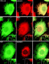
Peripheral structures containing both rbet1 and membrin are closely associated with, yet distinct from, ER tubules in fixed NRK cells. (Top row) Wild-type NRK cells grown at 37°C were fixed and stained for rbet1 (left) and membrin (middle) as described in Materials and Methods. Arrows represent peripheral structures positive for both proteins. Arrowheads represent peripheral structures positive for one, but not the other protein. (Middle row) Wild-type NRK cells grown at 37°C were fixed and stained for calnexin (left) and rbet1 (middle). (Bottom row) Wild-type NRK cells grown at 37°C were fixed and stained for Bip (left) and rbet1 (middle). Arrows in the middle and bottom rows point out peripheral rbet1- or membrin-positive structures very near, but distinct from, ER tubules. For Figs. 2, 4, and 5, the third column represents the merging of the first and second images. Bars, 10 μm (relative to objects in the first and second columns) and 4.3 μm (relative to insets). Insets show a higher magnification image from a similar cell.
Figure 4
Figure 4
Secretory cargo retained in the ER has access to a subset of peripheral structures containing rbet1 and membrin. (Top row) ts-G-GFP– transfected NRK cells grown at 40°C were fixed and the subcellular distribution of ts-G-GFP protein (left) was compared to the calnexin staining pattern (middle). (Middle row) ts-G-GFP–transfected NRK cells grown at 40°C were fixed and the subcellular distribution of ts-G-GFP (left) was compared to the rbet1 staining pattern (middle). (Bottom row) ts-G-GFP–transfected NRK cells grown at 40°C were fixed and the subcellular distribution of ts-G-GFP (left) was compared to the membrin staining pattern (middle). Right column represents an overlay of the previous two images in the row. Arrows point out peripheral rbet1- or membrin-containing structures that did not contain a significant amount of ts-G-GFP protein, although they were adjacent to ER tubules. The arrowheads demonstrate that some of the exclusive structures were not immediately adjacent to ER tubules, perhaps representing mobile VTCs en route to the Golgi complex. Bars: (frames) 10 μm; (inset) 4.3 μm.
Figure 5
Figure 5
GFP-labeled secretory cargo rapidly enters and accumulates in punctate rbet1- and membrin-containing structures when released from the ER. (Top row) ts-G-GFP–transfected NRK cells grown at 40°C and incubated for 5 min at 32°C were fixed and the subcellular distribution of ts-G-GFP (left) was compared to the calnexin staining pattern (middle). (Middle row) ts-G-GFP–transfected NRK cells grown at 40°C and incubated for 10 min at 32°C were fixed, and the subcellular distribution of ts-G-GFP (left) was compared to the rbet1 staining pattern (middle). (Bottom row) ts-G-GFP–transfected NRK cells grown at 40°C and incubated for 10 min at 32°C were fixed and the subcellular distribution of ts-G-GFP (left) was compared to the membrin staining pattern (middle). Right column respresents an overlay of the previous two images in the row. Arrows and arrowheads point out peripheral rbet1-containing structures that do not appear to overlap ER tubules, yet clearly contain the ts-G-GFP cargo. The arrowheads in the inset point to a structure enriched for both SNAREs and cargo that is clearly distinct from the ER, perhaps representing a mobile VTC en route to the Golgi complex. Bars: (frames) 10 μm; (insets) 4.3 μm.
Figure 3
Figure 3
COPII and rsec22b immunogold labeling of peripheral ER exit sites. Ultrathin cryosections of HepG2 cells double-immunogold–labeled for COPII (10 nm gold) and rsec22b (15 nm gold). (A) ER exit sites (arrowheads) with similar morphology are present near the nucleus, N, and close to the plasma membrane, P. (B) A higher magnification of a peripheral ER exit site showing the characteristic tubulovesicular morphology. M, mitochondrion. Bars, 200 nm.
Figure 6
Figure 6
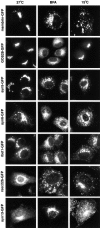
GFP-tagged SNAREs display a localization, sensitivity to BFA, and 15°C behavior similar to that of the endogenous proteins. Seven SNAREs were fused to GFP and images were captured at 37°C, in the presence of BFA for 1 h, or after 1 h at 15°C. The width of each panel is 56 μm.
Figure 7
Figure 7
rsec22b sites concentrate cargo that travel to the Golgi complex. (A) rsec22b-CFP labels peripheral sites. Time in minutes:seconds for A and B is indicated. (B) ts-G-YFP is localized in the ER and, upon release from the temperature block, becomes concentrated at preexisting rsec22b-CFP sites. (C) rsec22b-CFP labeling of peripheral sites. Time in minutes:seconds for C and D is indicated. (D) 5 min after release from the temperature block ts-G-GFP is depleted from rsec22b sites. Panel width is 6.2 μm.
Figure 8
Figure 8
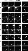
SNARE-GFP fusions undergo a variety of distinct movements. 1-s exposures are taken with 3 s between exposures. Symbols at the far right are used to represent the class of event. (A) Syntaxin 6-GFP; (B) rsec22b-GFP; (C) rsec22b-GFP; (D) lower magnification of C; (E) rsec22b-GFP; (F) rsec22b-GFP; (G) syntaxin 13-GFP; (H) syntaxin 13-GFP; and (I) rbet1-GFP. The width of a panel is 7.9 microns, except in panel D where the length is 22.2 μm.
Figure 9
Figure 9
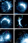
SNARE dynamics. (A) Sec22b-GFP–transfected cell with the pathways of individual organelles illustrated by different colors according to the key in Fig. 8. 1-s exposures were taken every 3 s. (B) rbet1-GFP. (C) syn5-GFP. (D) Membrin-GFP. An asterisk illustrates two structures that appear to associate with each other, whereas an open triangle marks two structures that appear to dissociate from each other. (E) Syntaxin 6-GFP is shown with 0.5-s exposures followed by 2.5 s of dark time. (F) Syntaxin 13-GFP. 0.5-s exposures followed by 1.5 s of dark time. Bars, 5 μm.
Figure 10
Figure 10
Overlapping and distinct SNARE trafficking pathways. Cotransfection of (A) rsec22b-CFP and (B) membrin-YFP reveals colocalizing structures (double arrow) and structures containing mostly membrin (arrowhead) or rsec22b (arrow). (C) A and B merged image. Moving structures that contain rsec22b and membrin appear as a double (yellow and cyan) spot because of the 1.25-s time difference between the exposures. Bars, 5 μm.

Similar articles

Cited by

References

    1. Advani RJ, Bae HR, Bock JB, Chao DS, Doung YC, Prekeris R, Yoo JS, Scheller RH. Seven novel mammalian SNARE proteins localize to distinct membrane compartments. J Biol Chem. 1998;273:10317–10324. - PubMed
    1. Alto MK, Ronne H, Keranen S. Yeast syntaxins Sso1p and Sso2p belong to a family of related membrane proteins that function in vesicular transport. EMBO (Eur Mol Biol Organ) J. 1993;12:4095–4104. - PMC - PubMed
    1. Balch WE, McCafferey JM, Plunter H, Farquhar MG. Vesicular stomatitis virus glycoprotein is sorted and concentrated during export from the endoplasmic reticulum. Cell. 1994;76:841–852. - PubMed
    1. Bennett MK, Scheller RH. The molecular machinery for secretion is conserved from yeast to neurons. Proc Natl Acad Sci USA. 1993;90:2559–2563. - PMC - PubMed
    1. Bergmann JE. Using temperature-sensitive mutants of VSV to study membrane protein biogenesis. Methods Cell Biol. 1989;32:85–110. - PubMed