. 1999 Mar 8;144(5):891-901.
doi: 10.1083/jcb.144.5.891.

Bid-induced conformational change of Bax is responsible for mitochondrial cytochrome c release during apoptosis

Affiliations

Bid-induced conformational change of Bax is responsible for mitochondrial cytochrome c release during apoptosis

S Desagher et al. J Cell Biol. .

Abstract

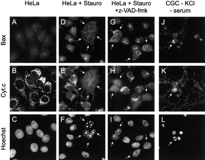
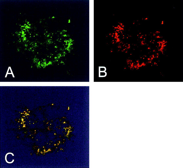
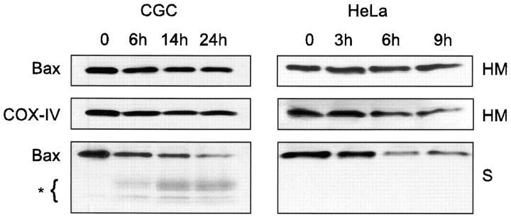
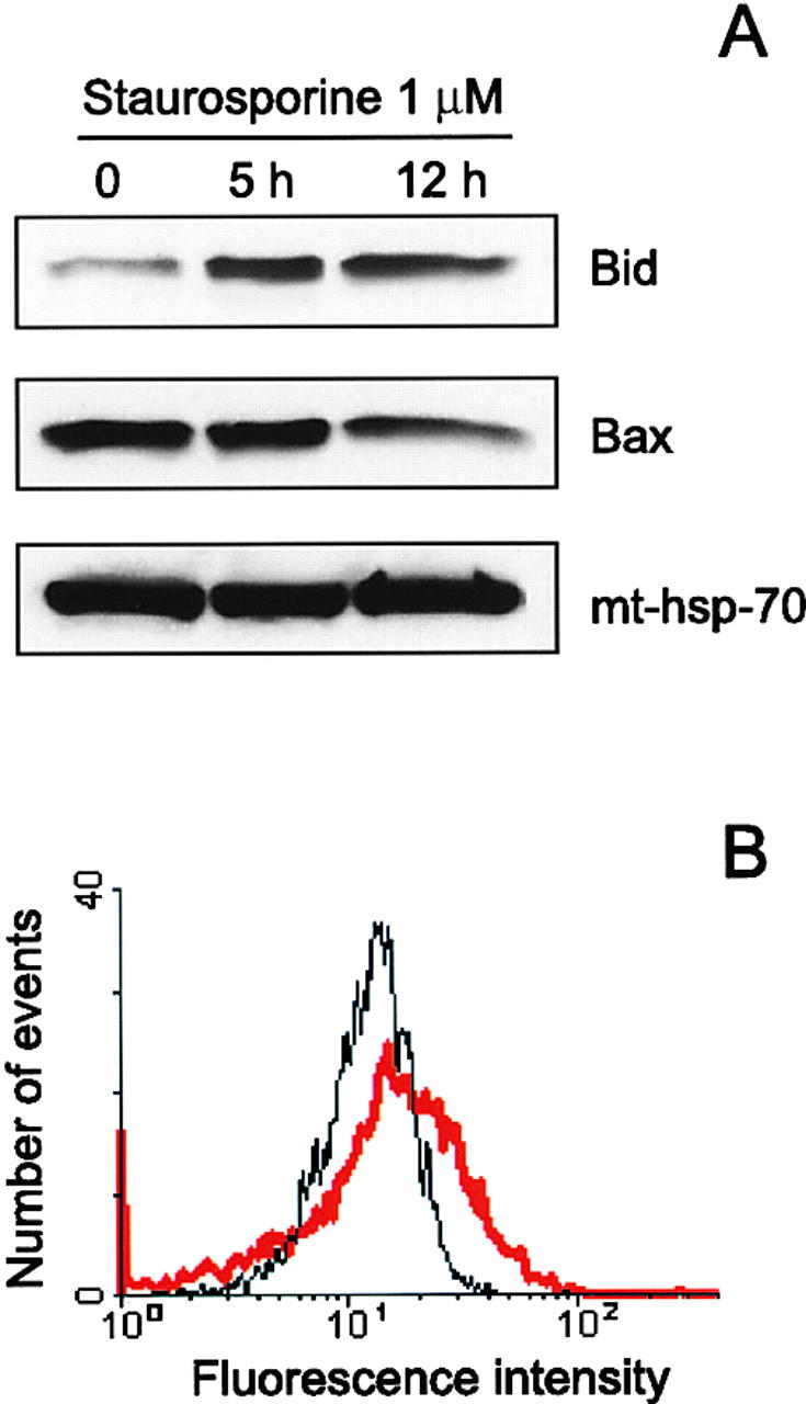
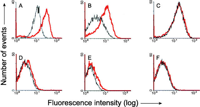
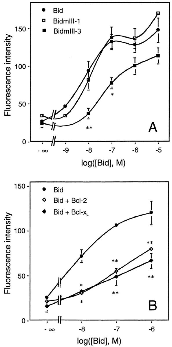
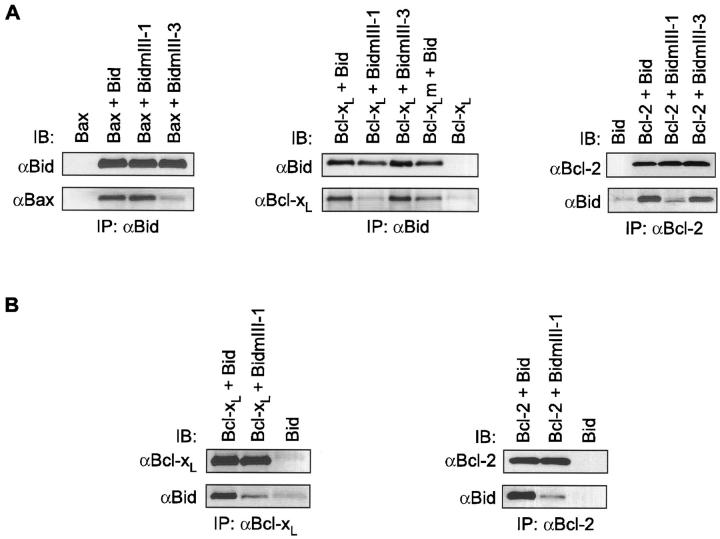
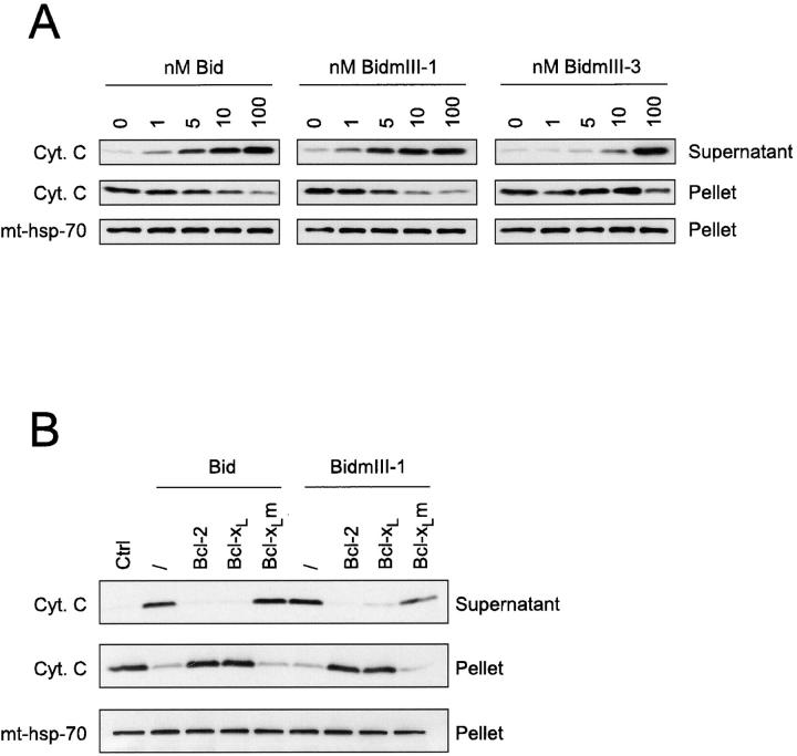
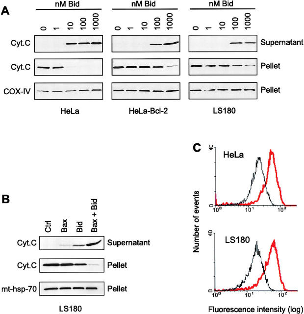
Here we report that in staurosporine-induced apoptosis of HeLa cells, Bid, a BH3 domain containing protein, translocates from the cytosol to mitochondria. This event is associated with a change in conformation of Bax which leads to the unmasking of its NH2-terminal domain and is accompanied by the release of cytochrome c from mitochondria. A similar finding is reported for cerebellar granule cells undergoing apoptosis induced by serum and potassium deprivation. The Bax-conformational change is prevented by Bcl-2 and Bcl-xL but not by caspase inhibitors. Using isolated mitochondria and various BH3 mutants of Bid, we demonstrate that direct binding of Bid to Bax is a prerequisite for Bax structural change and cytochrome c release. Bcl-xL can inhibit the effect of Bid by interacting directly with Bax. Moreover, using mitochondria from Bax-deficient tumor cell lines, we show that Bid- induced release of cytochrome c is negligible when Bid is added alone, but dramatically increased when Bid and Bax are added together. Taken together, our results suggest that, during certain types of apoptosis, Bid translocates to mitochondria and binds to Bax, leading to a change in conformation of Bax and to cytochrome c release from mitochondria.


Figures

Figure 1
Figure 1
Bax and cytochrome c immunostaining in cells undergoing apoptosis. (A–C) Control HeLa cells; (D–I) HeLa cells treated with 1 μM staurosporine for 4 h in the absence (D–F) or presence (G–I) of 100 μM z-VAD-fmk; (J–L) CGC cultured in a medium with low KCl (5 mM) and without serum for 7 h. Cells were double immunostained for Bax and cytochrome c and nuclei were visualized by Hoechst 33258 staining. A Bax pAb against amino acids 1–21 of human Bax (Upstate Biotechnology) was used in all cases. Arrows indicate Bax positive cells. Note the punctate Bax staining specifically in cells containing cytochrome c–depleted mitochondria. In CGC, in contrast to HeLa cells, the Bax immunostaining is seen only in neurons with a marginalized chromatin and not in neurons in the terminal phase of apoptosis (fragmented nucleus).
Figure 2
Figure 2
Bax staining appears in mitochondria. HeLa cells treated with 1 μM staurosporine for 2 h were double immunostained with antibodies against amino acids 11–30 of human Bax (Bax N-20 sc-493; Santa Cruz Biotechnology) and mt-hsp-70. Fluorescence microscopy was performed using a LSM 410 confocal microscope (Zeiss). The two channels are shown separately (A, anti-Bax; B, anti–mt-hsp-70) and merged (C).
Figure 3
Figure 3
Subcellular distribution of Bax in HeLa cells and CGC undergoing apoptosis. HeLa cells treated with 1 μM staurosporine for 3, 6, and 9 h and CGC cultured in a medium with a low KCl concentration (5 mM) and without serum for 6, 14, and 24 h were homogenized in isotonic buffer and separated into a soluble fraction (S) and an HM fraction enriched in mitochondria. The fractions (respectively, 30- and 15-μg proteins) were analyzed by Western blot with the pAb raised against amino acids 1–21 of human Bax (Upstate Biotechnology) and the monoclonal anti– COX-IV antibody.
Figure 4
Figure 4
Translocation of Bid to mitochondria during staurosporine-induced apoptosis of HeLa cells. (A) Mitochondria from HeLa cells before and after treatment with 1 μM staurosporine for 5 and 12 h were isolated on a sucrose gradient and analyzed for the presence of Bid and Bax by Western blotting. pAbs against full-length recombinant Bid (top) and to amino acids 1–21 of human Bax (middle) were used. Level of mt-hsp-70 (bottom) was used as a gel loading control. (B) Mitochondria from control HeLa cells (black curve) and HeLa cells treated with 1 μM staurosporine for 9 h (red curve) were isolated on a sucrose gradient, fixed, immunostained for Bid, and analyzed by flow cytometry.
Figure 5
Figure 5
Flow cytometric analysis of Bax immunofluorescence in mitochondria after exposure to Bid. Mitochondria from HeLa cells (A), HEK cells (B), Bcl-2–overexpressing HeLa cells (C), as well as from LoVo (D), DU145 (E), and LS180 (F) tumor cell lines, were isolated on a sucrose gradient and incubated for 15 min at 30°C in the presence or absence of 1 μM (A and C) or 8 μM recombinant Bid (B and D–F). After incubation, mitochondria were fixed and immunostained with the antibody to amino acids 1–21 of Bax (A and B, D–F) or with the antibody to amino acids 4–21 of human Bcl-2 (C). Immunostained mitochondria were analyzed by flow cytometry. Black curve: control. Red curve: after treatment with Bid.
Figure 7
Figure 7
Dose–response curve of Bid-induced Bax immunoreactivity. (A) Effects of BH3 mutations in Bid. Mitochondria from HeLa cells were isolated on a sucrose gradient and incubated for 15 min at 30°C with increasing concentrations of recombinant wild-type Bid or BidmIII-1 and BidmIII-3 mutants. Mitochondria were then fixed, immunostained with the antibody raised against amino acids 1–21 of Bax, and analyzed by flow cytometry. (B) Inhibition of Bid effect by Bcl-2 and Bcl-xL. Mitochondria isolated from HeLa cells were incubated with increasing concentrations of recombinant Bid for 15 min at 30°C in the presence or absence of 1 μM recombinant Bcl-2 or Bcl-xL before fixation, Bax immunostaining, and FACS® analysis. Results are mean ± SEM of three independent experiments, each performed in duplicate. *P < 0.05; **P < 0.01; significantly different from corresponding values obtained with wild-type Bid (ANOVA followed by Bonferroni's test).
Figure 6
Figure 6
Interactions between BH3 mutants of Bid and Bcl-2 family proteins in vitro. (A) 100 nM of Bid, BidmIII-1, or BidmIII-3 was incubated in PBS with 100 nM of Bax, Bcl-xL, Bcl-xLm, or Bcl-2 for 30 min on ice. Recombinant proteins were then immunoprecipitated (IP) with the pAb against Bid or a mAb against Bcl-2 (Genosys) and separated by SDS-PAGE. Immunoblottings (IB) were performed using the pAb against Bid, an mAb against Bax (Genzyme), a pAb against Bcl-x (Transduction Laboratories), or a pAb against Bcl-2 (sc-492). (B) 100 nM of Bid or BidmIII-1 was incubated with 1 μM of Bcl-xL or Bcl-2 for 30 min on ice, followed by steps described in A. The same antibodies were used for immunoprecipitation and immunoblotting. In addition, the polyclonal anti–Bcl-x antibody from Santa Cruz Biotechnology (sc-634) was used to precipitate Bcl-xL.
Figure 8
Figure 8
Bid-induced cytochrome c release from mitochondria. (A) Mitochondria from HeLa cells were isolated on a sucrose gradient, incubated for 15 min at 30°C with increasing concentrations of recombinant Bid as indicated. The incubation mix was centrifuged and the supernatants and pellets were analyzed for cytochrome c content by Western blot. mt-hsp-70 was used as a gel loading control. (B) Mitochondria from HeLa cells isolated on a sucrose gradient were incubated for 15 min at 30°C with 100 nM of wild-type Bid in the presence or absence of 1 μM of Bcl-2, Bcl-xL, or a mutant of Bcl-xL in which Gly 138 was replaced by an alanine (Bcl-xLm). Mitochondria were then centrifuged and the cytochrome c content of both the pellet and the supernatant of each sample was estimated by Western blotting. Equal loading of the mitochondrial pellet was confirmed with the antibody to mt-hsp-70.
Figure 9
Figure 9
Involvement of Bax and Bcl-2 in the Bid-induced release of cytochrome c from mitochondria. (A) Mitochondria were isolated on a sucrose gradient from HeLa cells, Bcl-2–overexpressing HeLa cells, and from the Bax-deficient colon tumor cells LS180. They were incubated for 15 min at 30°C in the presence of increasing concentrations of recombinant Bid and the contents of cytochrome c of both the mitochondrial pellets and the supernatants were estimated by Western blot. Equal loading of the mitochondrial pellet was confirmed with the monoclonal anti–COX-IV antibody. (B) Mitochondria from LS180 cells were incubated for 15 min at 30°C with 5 μM recombinant Bax lacking 20 amino acids at the COOH terminus in the presence or absence of 50 nM recombinant Bid. Cytochrome c was detected by Western blot in the mitochondrial pellets and the supernatants. Equal loading of the mitochondrial pellet was confirmed with the antibody against mt-hsp-70. (C) Flow cytometric analysis of Bak immunostaining of mitochondria isolated from HeLa and LS180 cells. Mitochondria from both cell types were isolated on a sucrose gradient and incubated with 1 μM recombinant Bid for 15 min at 30°C before fixation, immunostaining with an antibody raised against NH2-terminal domain of Bak (amino acids 1–52; Calbiochem), and FACS® analysis. Black curve: control. Red curve: 1 μM Bid.

References

    1. Antonsson B, Conti F, Ciavatta AM, Montessuit S, Lewis S, Martinou I, Bernasconi L, Bernard A, Mermod J-J, Mazzei G, et al. Inhibition of Bax channel-forming activity by Bcl-2. Science. 1997;277:370–372. - PubMed
    1. Aritomi M, Kunishima N, Inohara N, Ishibashi Y, Ohta S, Morikawa K. Crystal structure of rat Bcl-xL . J Biol Chem. 1997;272:27886–27892. - PubMed
    1. Bossy-Wetzel E, Newmeyer DD, Green DR. Mitochondrial cytochrome c release in apoptosis occurs upstream of DEVD-specific caspase activation and independently of mitochondrial transmembrane depolarization. EMBO (Eur Mol Biol Organ) J. 1998;17:37–49. - PMC - PubMed
    1. Bradford MM. A rapid and sensitive method for the quantitation of microgram quantities of protein utilizing the principle of protein dye binding. Anal Biochem. 1976;72:248–254. - PubMed
    1. Cheng EH-Y, Levine B, Boise LH, Thompson CB, Hardwick JM. Bax-independent inhibition of apoptosis by Bcl-xL . Nature. 1996;379:554–556. - PubMed

Publication types

MeSH terms