. 1999 Mar 8;144(5):989-1000.
doi: 10.1083/jcb.144.5.989.

Specific myosin heavy chain mutations suppress troponin I defects in Drosophila muscles

Affiliations

Specific myosin heavy chain mutations suppress troponin I defects in Drosophila muscles

W A Kronert et al. J Cell Biol. .

Abstract

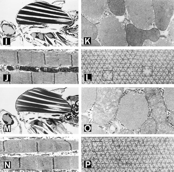
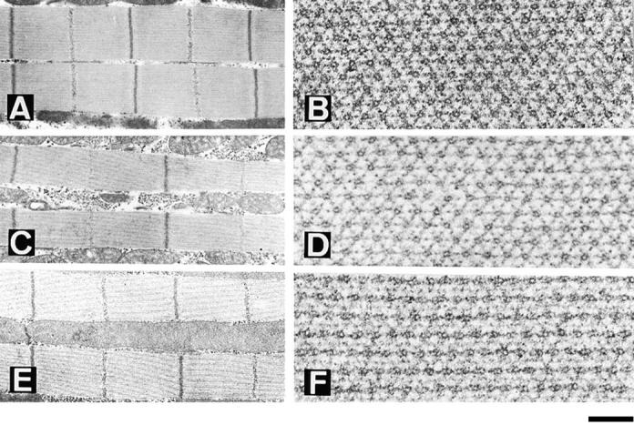
We show that specific mutations in the head of the thick filament molecule myosin heavy chain prevent a degenerative muscle syndrome resulting from the hdp2 mutation in the thin filament protein troponin I. One mutation deletes eight residues from the actin binding loop of myosin, while a second affects a residue at the base of this loop. Two other mutations affect amino acids near the site of nucleotide entry and exit in the motor domain. We document the degree of phenotypic rescue each suppressor permits and show that other point mutations in myosin, as well as null mutations, fail to suppress the hdp2 phenotype. We discuss mechanisms by which the hdp2 phenotypes are suppressed and conclude that the specific residues we identified in myosin are important in regulating thick and thin filament interactions. This in vivo approach to dissecting the contractile cycle defines novel molecular processes that may be difficult to uncover by biochemical and structural analysis. Our study illustrates how expression of genetic defects are dependent upon genetic background, and therefore could have implications for understanding gene interactions in human disease.


Figures

Figure 1
Figure 1
Normal and heldup2 mutant dorsolongitudinal muscles (DLM). (A) Diagram showing the six (a–f) DLM fibers in a sagittal view. Anterior is to the left, and dorsal is up. (B) Detail of a fibril showing a sarcomere between Z bands and including an M line. (C) Transverse view of a fibril showing the 1:6 array of thick to thin filaments. The square box has one thick filament in the center and six surrounding thin filaments in a hexagonal pattern. (D) Sagittal section of a hdp2 male. Note the remnants of the six DLMs near their attachment sites. (E) Hypercontracted sarcomeres in which the M line is no longer detected. (F) Cross section of a mutant muscle in which only thick filaments are visible. Bar: (A) 250 μm; (B and E) 750 nm; (C and F) 120 nm; (D) 240 μm.
Figure 2
Figure 2
Locations of mutations on the three-dimensional structure of the myosin head. The figure depicts a stereo-pair image of the atomic resolution structure of chicken myosin S1 (Rayment et al., 1993b) with regions encoded by Drosophila alternative exons and Drosophila Mhc mutations superimposed. The backbone of the heavy chain is yellow, the essential light chain is green, and the regulatory light chain is red. The location of the β-phosphate group of ATP is shown as a red sphere, with the reactive thiols depicted as tricolored residues. The Drosophila MHC regions encoded by alternative exons are purple (exon 3), light blue (exon 7), dark blue (exon 9), and tan (exon 11; Bernstein and Milligan, 1997). Two hdp2 suppressor mutations are at the actin binding loop. Mutation D1 affects residue 625 at the base of the loop while mutation D62 is an 8 amino acid deletion in the loop itself. The loop is not visible in the structure due to its flexible nature, but its ends are located behind residue 625 and at the terminus of a long helical structure just to its right. Mutation D45 is at residue 261, on the surface of the molecule. This residue is just to the right of the two free ends of the molecule representing the flexible loop at the lip of the nucleotide binding pocket. The loop itself is not visible in the structure. Mutation D41 affects the lip of the nucleotide pocket by causing the substitution of exon 7 alternative exons (light blue). Residue 200, which is mutated in Mhc5, is also in this general vicinity of the molecule and acts to enhance the effects of hdp2. A mutation in the regulatory light chain binding domain at residue 832 (Mhc8) enhances the effects of the hdp2 allele to a lesser extent. Graphic produced in collaboration with Dr. Ronald Milligan (The Scripps Research Institute) using the Molscript program.
Figure 3
Figure 3
In situ hybridization analysis of Mhc mRNA accumulation in wild-type and D41 Mhc mutant. D41 mutation creates a splice junction-like sequence resulting in misregulation of alternative splicing of the exon 7 series. Probes specific to alternative versions of exon 7 show that exon 7d is used in wild-type indirect flight muscles, but replaced with exon 7c in D41. Panels are brightfield micrographs of parasagittal sections of thoraces (anterior is left, dorsal is up). (A) Wild-type male probed with exon 7c, showing no hybridization to the indirect flight muscles that comprise the bulk of the upper thoracic region. (B) Wild-type male probed with exon 7d, showing strong hybridization to the indirect flight muscles. (C) hdp2/Y;D41/Mhc10 males probed with exon 7c, showing strong hybridization to the indirect flight muscles. (D) hdp2/Y;D41/Mhc10 males probed with exon 7d showing failure of the indirect flight muscles to hybridize with this probe.
Figure 4
Figure 4
Suppression of the troponin I mutation hdp2 by the D-series Mhc mutations. D1 (A–D) and D62 (E–H) affect the actin-binding loop of MHC. (A) Sagittal, slightly tilted, view of a hdp2;D1/+ male. Note the almost normal appearance of DLM fibers a–d but the collapse of muscles e and f (arrow- head). (B) Detail of two fibrils. Note the restoration of the M line in the sarcomeres. (C) Cross section of a suppressed muscle. m = mitochondrion. (D) Detail of the thick–thin filament array. Some structural failures such as the absence of a thick filament (arrow- head) or an additional thin filament (arrow) can be identified. (E) Sagittal view of a hdp2;D62/+ male. Note the persistence of gross structural defects on muscles e and f near the posterior attachment site. (F) Detail of fibrils showing virtually normal sarcomeres. (G) Cross section showing a near normal fibril. The apparent disorganization indicated by an arrowhead is an artifact of preparation, found occasionally in wild-type. (H) Detail of the filament array. D41 (I–L) and D45 (M–P) are mutations near the nucleotide entry site in MHC. (I) Sagittal view of a hdp2;D41/+ male. There are persistent structural defects towards the posterior side of muscles e and f. (J) Detail of the fibrils. Note the incomplete definition of Z and M bands towards the edge of the fibril (double arrows). (K) Cross section of fibrils showing the disorganized array at the periphery (star). (L) Detail of the filament array. Occasionally, thick filaments are absent or a thin filament substitutes for thick (squares). (M) Sagittal section of an hdp2;D45/+ male. As with all other suppressors, gross structural defects persist toward the posterior site of e and f muscle, although, in this case, d is also visibly affected. (N) Sarcomeres with incomplete restoration of Z and M lines. (O) Cross view of fibrils. (P) Detail of filament array showing a double thick filament (square). Bar: (A, E, I, and M) 330 μm; (B, F, J, and N) 775 nm; (C, G, K, and O) 430 nm; (D, H, L, and P) 144 nm.
Figure 4
Figure 4
Suppression of the troponin I mutation hdp2 by the D-series Mhc mutations. D1 (A–D) and D62 (E–H) affect the actin-binding loop of MHC. (A) Sagittal, slightly tilted, view of a hdp2;D1/+ male. Note the almost normal appearance of DLM fibers a–d but the collapse of muscles e and f (arrow- head). (B) Detail of two fibrils. Note the restoration of the M line in the sarcomeres. (C) Cross section of a suppressed muscle. m = mitochondrion. (D) Detail of the thick–thin filament array. Some structural failures such as the absence of a thick filament (arrow- head) or an additional thin filament (arrow) can be identified. (E) Sagittal view of a hdp2;D62/+ male. Note the persistence of gross structural defects on muscles e and f near the posterior attachment site. (F) Detail of fibrils showing virtually normal sarcomeres. (G) Cross section showing a near normal fibril. The apparent disorganization indicated by an arrowhead is an artifact of preparation, found occasionally in wild-type. (H) Detail of the filament array. D41 (I–L) and D45 (M–P) are mutations near the nucleotide entry site in MHC. (I) Sagittal view of a hdp2;D41/+ male. There are persistent structural defects towards the posterior side of muscles e and f. (J) Detail of the fibrils. Note the incomplete definition of Z and M bands towards the edge of the fibril (double arrows). (K) Cross section of fibrils showing the disorganized array at the periphery (star). (L) Detail of the filament array. Occasionally, thick filaments are absent or a thin filament substitutes for thick (squares). (M) Sagittal section of an hdp2;D45/+ male. As with all other suppressors, gross structural defects persist toward the posterior site of e and f muscle, although, in this case, d is also visibly affected. (N) Sarcomeres with incomplete restoration of Z and M lines. (O) Cross view of fibrils. (P) Detail of filament array showing a double thick filament (square). Bar: (A, E, I, and M) 330 μm; (B, F, J, and N) 775 nm; (C, G, K, and O) 430 nm; (D, H, L, and P) 144 nm.
Figure 5
Figure 5
Double suppressor combinations. Longitudinal (A, C, and E) and transverse (B, D, and F) views of D1/ D45 (A and B), D1/D41 (C and D) and D1/D62 (E and F) dorsolongitudinal muscles in males carrying the hdp2 mutation. Note the improved restoration of muscle structure in cases of D41 and D45 combinations with D1 suggesting independent and additive mechanisms of suppression. In the case of the D1/ D62 combination, although there is an additional improvement in the restoration of sarcomere and filament array, there is a new structural feature: the frequent assembly of thick filaments in pairs. Bar: (A, C, and E) 775 nm; (B, D, and F) 144 nm.

Similar articles

Cited by

References

    1. al-Khayat HA, Yagi N, Squire JM. Structural changes in actin-tropomyosin during muscle regulation: computer modelling of low-angle X-ray diffraction data. J Mol Biol. 1995;252:611–632. - PubMed
    1. Barbas JA, Galceran J, Torroja L, Prado A, Ferrús A. Abnormal muscle development in the heldup3 mutant of Drosophila melanogasteris caused by a splicing defect affecting selected troponin I isoforms. Mol Cell Biol. 1993;13:1433–1439. - PMC - PubMed
    1. Beall CJ, Fyrberg E. Muscle abnormalities in Drosophila melanogasterheldup mutants are caused by missing or aberrant troponin-I isoforms. J Cell Biol. 1991;114:941–951. - PMC - PubMed
    1. Bernstein SI, Milligan RA. Fine tuning a molecular motor: the location of alternative domains in the Drosophilamyosin head. J Mol Biol. 1997;271:1–6. - PubMed
    1. Collier VL, Kronert WA, O'Donnell PT, Edwards KA, Bernstein SI. Alternative myosin hinge regions are utilized in a tissue-specific fashion that correlates with muscle contraction speed. Genes Dev. 1990;4:885–895. - PubMed

Publication types