. 1999 Apr 1;19(7):2658-64.
doi: 10.1523/JNEUROSCI.19-07-02658.1999.

Stimulus-dependent translocation of kappa opioid receptors to the plasma membrane

Affiliations

Stimulus-dependent translocation of kappa opioid receptors to the plasma membrane

S J Shuster et al. J Neurosci. .

Abstract

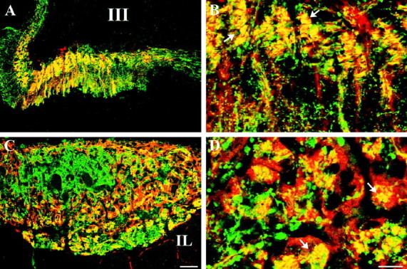
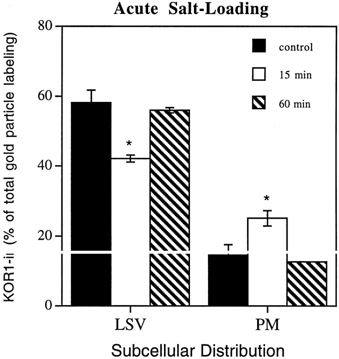
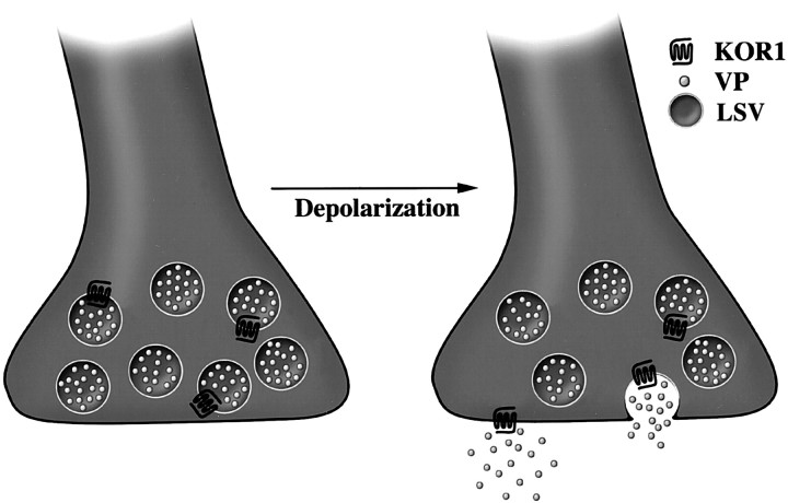
We examined the cellular and subcellular distribution of the cloned kappa opioid receptor (KOR1) and its trafficking to the presynaptic plasma membrane in vasopressin magnocellular neurosecretory neurons. We used immunohistochemistry to show that KOR1 immunoreactivity (IR) colocalized with vasopressin-containing cell bodies, axons, and axon terminals within the posterior pituitary. Ultrastructural analysis revealed that a major fraction of KOR1-IR was associated with the membrane of peptide-containing large secretory vesicles. KOR1-IR was rarely associated with the plasma membrane in unstimulated nerve terminals within the posterior pituitary. A physiological stimulus (salt-loading) that elicits vasopressin release also caused KOR1-IR to translocate from these vesicles to the plasma membrane. After stimulation, there was a significant decrease in KOR1-IR associated with peptide-containing vesicles and a significant increase in KOR1-IR associated with the plasma membrane. This stimulus-dependent translocation of receptors to the presynaptic plasma membrane provides a novel mechanism for regulation of transmitter release.


Figures

Fig. 1.
Fig. 1.
KOR1- and VPNP-IR colocalize in the cell bodies of hypothalamic MNN. Confocal micrographs of KOR1 (A,C) and VPNP (B, D) double-labeled single sections. A, B, Single section of rat paraventricular nucleus double-labeled for KOR1-IR (A) and VPNP-IR (B). Examples of KOR1 and VPNP colocalization within cell bodies are indicated by arrows.C, D, Single section of rat supraoptic nucleus double-labeled for KOR1-IR (C) and VPNP-IR (D). Examples of KOR1 and VPNP colocalization within cell bodies are indicated byarrows. Scale bar: A–D, 100 μm.
Fig. 2.
Fig. 2.
A large portion of KOR1- and VPNP-IR colocalize in the same structures in the axons of the median eminence and nerve terminals within the neural lobe of the pituitary. Confocal micrographs of KOR1-IR (red) and VPNP-IR (green) in double-labeled single sections of rat median eminence (A, B) and posterior pituitary (C, D). Instances of colocalization are indicated by yellow (andarrows), created by the digital merging ofred (KOR-IR) and green (VPNP-IR).A, Low-magnification image of median eminence showing colocalization of KOR1- and VPNP-IR within the internal layer of the median eminence and scattered fibers in the external layer.B, High-magnification image showing colocalization of KOR1- and VPNP-IR in discrete puncta in a subset of fibers within the internal layer of the median eminence. C, Low-magnification image of nerve terminals within the neural lobe that are positive for both KOR1- and VPNP-IR. D, High-magnification image showing that KOR1- and VPNP-IR are colocalized within a subpopulation of the nerve terminals. Scale bars: (inC) A, C, 50 μm; (inD) B, D, 30 μm.III, Third ventricle; IL, intermediate lobe of the pituitary.
Fig. 3.
Fig. 3.
KOR1-IR is associated with the membrane of large secretory vesicles containing VPNP-IR. Transmission electron microscopy micrographs of postembedding–immunogold staining for KOR1 (15 nm gold) and VPNP (5 nm gold) within the neural lobe. Serial sections single-labeled with anti-KOR1 (A) and anti-VPNP (B). The same vesicle (small arrows) is labeled with KOR1- and VPNP-IR in both sections (A, B). C, Single section double-labeled with anti-KOR1 (15 nm) and anti-VPNP (5 nm) also showing KOR1- and VPNP-IR colocalized in the same large secretory vesicle (large arrow). Single-labeled sections showing KOR1-IR on the membrane of a large secretory vesicle (D) and the plasma membrane (E). Scale bars: (inB) A, B, 250 nm;C, 100 nm; (in E) D,E, 100 nm.
Fig. 4.
Fig. 4.
Subcellular distribution of KOR1 in the nerve terminals of rat posterior pituitary. The graph shows the summary of the quantification of immunogold particles representing KOR1-IR, expressed as percentage of total KOR1-IR. Subcellular compartments were defined as large secretory vesicles (LSV), plasma membrane (PM), cytoplasm (CYTO), and synaptic-like microvesicles (SLMV). Large secretory vesicles, 62.5 ± 1.8%; plasma membrane, 10.8 ± 1.2%; cytoplasm, 17 ± 2.1%; synaptic-like microvesicles, 10.7 ± 1.8%. Error bars indicate ±SEM.
Fig. 5.
Fig. 5.
KOR1-IR translocates to the plasma membrane from large secretory vesicles in a stimulus-dependent manner. Experimental animals (n = 3) were treated with an intraperitoneal injection of hypertonic saline 15 or 60 min before perfusion fixation. In control animals (n = 3), a needle was inserted and withdrawn without the delivery of saline. The graph shows the summary of the quantification of immunogold particles representing KOR1-IR, expressed as percentage of total KOR1-IR. The values shown represent the mean ± SEM. Gold particles were counted in 464 nerve terminals. Control: large secretory vesicles, 58.2 ± 3.6%; plasma membrane, 14.6 ± 3.0%; 15 min stimulation: large secretory vesicles, 42.1 ± 1.0%; plasma membrane, 25.2 ± 2.2%; 60 min stimulation: large secretory vesicles, 56.0 ± 0.7%; plasma membrane, 12.8 ± 0.2%. *p < 0.05. Error bars indicate ±SEM.
Fig. 6.
Fig. 6.
Schematic illustration showing translocation of presynaptic KOR1 from its transport vesicle to the plasma membrane. In the nerve terminals of the neural lobe, KOR1 appears to be transported in vesicles containing vasopressin (VP). Conditions that cause depolarization and release of neurohormone appear to cause KOR1 to be inserted into the plasma membrane, giving the receptor access to its ligand to transduce a signal across the plasma membrane.

References

    1. Adams JC. Biotin amplification of biotin and horseradish peroxidase signals in histochemical stains. J Histochem Cytochem. 1992;40:1457–1463. - PubMed
    1. Arvidsson U, Riedl M, Chakrabarti S, Vulchanova L, Lee J-H, Nakano AH, Lin X, Loh HH, Law P-Y, Wessendorf MW, Elde R. The κ-opioid receptor (KOR1) is primarily postsynaptic: combined immunohistochemical localization of the receptor and endogenous opioids. Proc Natl Acad Sci USA. 1995;92:5062–5066. - PMC - PubMed
    1. Berghorn KA, Bonnett JH, Hoffman GE. cFos immunoreactivity is enhanced with biotin amplification. J Histochem Cytochem. 1994;42:1635–1642. - PubMed
    1. Boersma CJC, Sonnemans MA, Van Leeuwen FW. Immunoelectron microscopic demonstration of oxytocin and vasopressin in pituicytes and in nerve terminals forming synaptoid contacts with pituicytes in the rat neural lobe. Brain Res. 1993;611:117–129. - PubMed
    1. Boersma CJC, Pool CW, Van Heerikhuize J, Van Leeuwen FW. Characterization of opioid binding sites in the neural and intermediate lobe of the rat pituitary gland by quantitative receptor autoradiography. J Neuroendocrinol. 1994;6:47–56. - PubMed

Publication types

LinkOut - more resources