

Interleukin-1beta in immune cells of the abdominal vagus nerve: a link between the immune and nervous systems?
- PMID: 10087091
- PMCID: PMC6786076
- DOI: 10.1523/JNEUROSCI.19-07-02799.1999
Interleukin-1beta in immune cells of the abdominal vagus nerve: a link between the immune and nervous systems?
Abstract
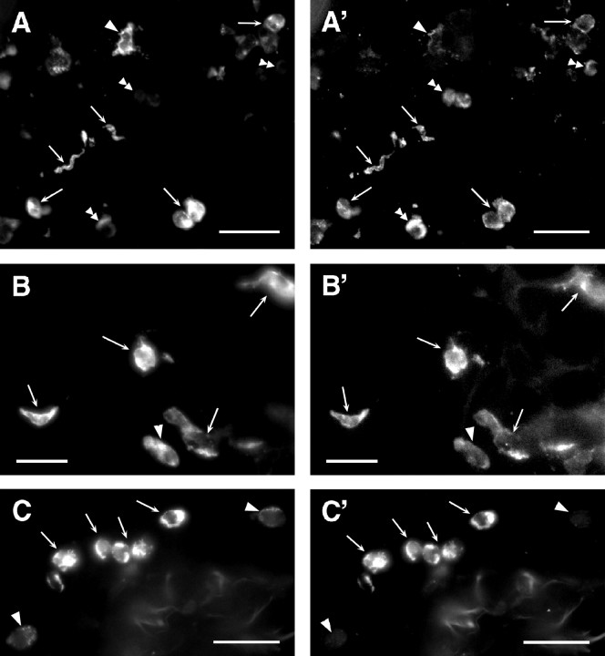
Intraperitoneal administration of the cytokine interleukin-1beta (IL-1beta) induces brain-mediated sickness symptoms that can be blocked by subdiaphragmatic vagotomy. Intraperitoneal IL-1beta also induces expression of the activation marker c-fos in vagal primary afferent neurons, suggesting that IL-1beta is a key component of vagally mediated immune-to-brain communication. The cellular sources of IL-1beta activating the vagus are unknown, but may reside in either blood or in the vagus nerve itself. We assayed IL-1beta protein after intraperitoneal endotoxin [lipopolysaccharide (LPS)] injection in abdominal vagus nerve, using both an ELISA and immunohistochemistry, and in blood plasma using ELISA. IL-1beta levels in abdominal vagus nerve increased by 45 min after LPS administration and were robust by 60 min. Plasma IL-1beta levels increased by 60 min, whereas little IL-1beta was detected in cervical vagus or sciatic nerve. IL-1beta-immunoreactivity (IR) was expressed in dendritic cells and macrophages within connective tissues associated with the abdominal vagus by 45 min after intraperitoneal LPS injection. By 60 min, some immune cells located within the nerve and vagal paraganglia also expressed IL-1beta-IR. Thus, intraperitoneal LPS induced IL-1beta protein within the vagus in a time-frame consistent with signaling of immune activation. These results suggest a novel mechanism by which IL-1beta may serve as a molecular link between the immune system and vagus nerve, and thus the CNS.
Figures


References
-
- Banchereau J, Steinman RM. Dendritic cells and the control of immunity. Nature. 1998;392:425–252. - PubMed
-
- Berthoud H-R, Kressel M, Neuhuber WL. Vagal afferent innervation of rat abdominal paraganglia as revealed by anterograde DiI-tracing and confocal microscopy. Acta Anat. 1995;152:127–132. - PubMed
-
- Bradford MM. A rapid and sensitive method for quantification of microgram quantities of protein utilizing the principle of protein-dye binding. Anal Biochem. 1976;72:248–254. - PubMed
-
- Bret-Dibat JL, Bluthé R-M, Kent S, Kelley K, Dantzer R. Lipopolysaccharide and interleukin-1 depress food-motivated behavior in mice by a vagal-mediated mechanism. Brain Behav Immun. 1995;9:242–246. - PubMed
-
- Carlson DE. Adrenocorticotropin correlates strongly with endotoxemia after intravenous but not after intraperitoneal inoculations of E. coli. Shock. 1997;7:65–69. - PubMed
Publication types
MeSH terms
Substances
Grants and funding
LinkOut - more resources
Full Text Sources