. 1999 Mar 22;144(6):1113-22.
doi: 10.1083/jcb.144.6.1113.

Dynamics of myoblast transplantation reveal a discrete minority of precursors with stem cell-like properties as the myogenic source

Affiliations

Dynamics of myoblast transplantation reveal a discrete minority of precursors with stem cell-like properties as the myogenic source

J R Beauchamp et al. J Cell Biol. .

Abstract

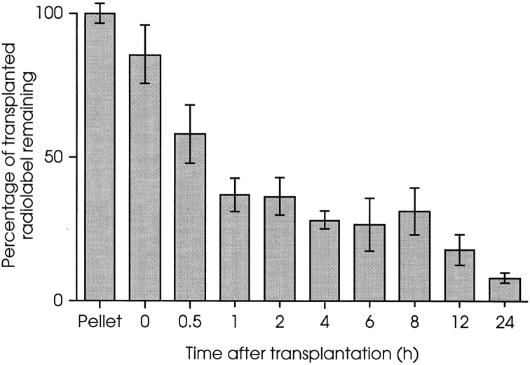
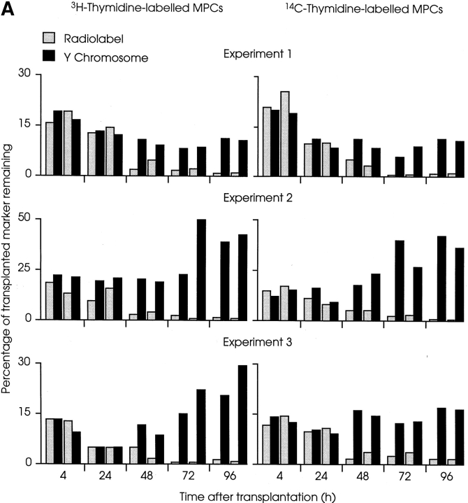
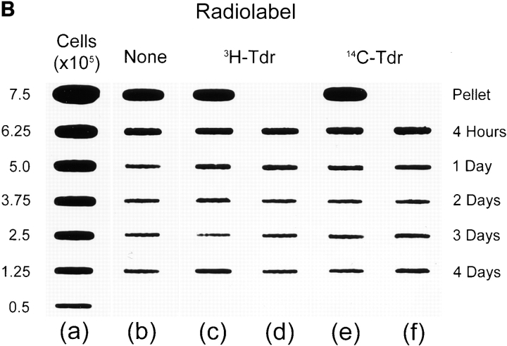
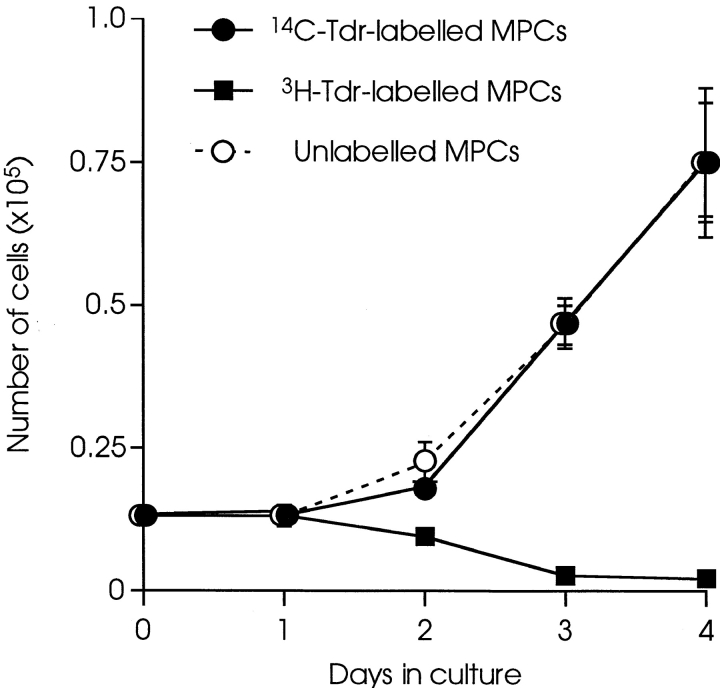
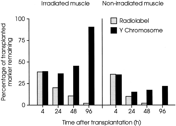
Myoblasts, the precursors of skeletal muscle fibers, can be induced to withdraw from the cell cycle and differentiate in vitro. Recent studies have also identified undifferentiated subpopulations that can self-renew and generate myogenic cells (Baroffio, A., M. Hamann, L. Bernheim, M.-L. Bochaton-Pillat, G. Gabbiani, and C.R. Bader. 1996. Differentiation. 60:47-57; Yoshida, N., S. Yoshida, K. Koishi, K. Masuda, and Y. Nabeshima. 1998. J. Cell Sci. 111:769-779). Cultured myoblasts can also differentiate and contribute to repair and new muscle formation in vivo, a capacity exploited in attempts to develop myoblast transplantation (MT) for genetic modification of adult muscle. Our studies of the dynamics of MT demonstrate that cultures of myoblasts contain distinct subpopulations defined by their behavior in vitro and divergent responses to grafting. By comparing a genomic and a semiconserved marker, we have followed the fate of myoblasts transplanted into muscles of dystrophic mice, finding that the majority of the grafted cells quickly die and only a minority are responsible for new muscle formation. This minority is behaviorally distinct, slowly dividing in tissue culture, but rapidly proliferative after grafting, suggesting a subpopulation with stem cell-like characteristics.


Figures

Figure 2
Figure 2
Biphasic loss of donor MPCs from irradiated host muscles during the initial 24 h after transplantation. [14C]Thymidine– labeled H-2Kbclone 18 MPCs were injected into irradiated left TA muscles of nude mdx mice. Each muscle received 5 × 10⁵ cells and at the times indicated, amount of 14C present in host muscles was measured. Less than 10% of the injected radiolabel remained 24 h after MT. 60% was lost during the first 60 min and a second event, initiated ≥8 h after MT, resulted in the subsequent disappearance of most of the remaining label. Results are presented as percentages of 14C present in 5 × 10⁵ labeled MPCs, and each time point is the mean value from four muscles ± 1 SD. Muscles taken at 0 h were removed as quickly as possible after MT (i.e., <5 min) and contained ∼85% of the injected label.
Figure 1
Figure 1
Loss and proliferation of MPCs following myoblast transplantation into irradiated host muscle. (A) Irradiated left TA muscles of female nude mdx mice were transplanted with 5 × 10⁵ radiolabeled male H-2Kbclone 18 MPCs. At the times indicated, mice were killed and amounts of Y chromosome (filled columns) and radiolabel (open columns) were measured and are presented as percentages. In each experiment, most donor MPCs disappeared from host muscles during the initial 24 h after MT, as shown by a coincident loss of the majority of donor cell markers. During the subsequent 3 d, amounts of Y chromosome revealed marked proliferation of surviving cells, despite the continued, progressive disappearance of the radiolabeled population. In each experiment shown, four mice were killed at each time point, two had received [3H]thymidine–labeled MPCs (left), and two had been transplanted with [14C]thymidine–labeled MPCs (right). Each pair of columns represents percentages of radiolabel and Y chromosome remaining in an individual muscle. (B) Representative slot blot of DNA extracted from irradiated female nude mdx mouse muscles injected with unlabeled or radiolabeled male MPCs, probed for Y chromosome. (a) Series of control samples prepared from noninjected female muscles combined with 7.5–0.5 × 10⁵ male MPCs; (b) DNA extracted from muscles injected with 5 × 10⁵ unlabeled cells; (c and d) DNA extracted from muscles injected with the same number of [3H]thymidine–labeled MPCs; and (e and f) DNA from muscles injected with [14C]thymidine–labeled cells. The time at which each muscle was harvested after MT is shown at the right of the figure. The top slots in b, c, and e contain DNA extracted from representative cell pellets containing 5 × 10⁵ unlabeled or labeled MPCs. The signal from each slot was measured using a PhosphoImager and values obtained from a were used to construct a linear standard curve of signal against number of donor cells (data not shown). Signals obtained from injected muscles were within the linear range of this system, which is maintained with as little as 1 ng of target DNA (equivalent to ∼200 genome copies; Beauchamp et al., 1997). Values obtained from this blot are presented in A, experiment 1.
Figure 1
Figure 1
Loss and proliferation of MPCs following myoblast transplantation into irradiated host muscle. (A) Irradiated left TA muscles of female nude mdx mice were transplanted with 5 × 10⁵ radiolabeled male H-2Kbclone 18 MPCs. At the times indicated, mice were killed and amounts of Y chromosome (filled columns) and radiolabel (open columns) were measured and are presented as percentages. In each experiment, most donor MPCs disappeared from host muscles during the initial 24 h after MT, as shown by a coincident loss of the majority of donor cell markers. During the subsequent 3 d, amounts of Y chromosome revealed marked proliferation of surviving cells, despite the continued, progressive disappearance of the radiolabeled population. In each experiment shown, four mice were killed at each time point, two had received [3H]thymidine–labeled MPCs (left), and two had been transplanted with [14C]thymidine–labeled MPCs (right). Each pair of columns represents percentages of radiolabel and Y chromosome remaining in an individual muscle. (B) Representative slot blot of DNA extracted from irradiated female nude mdx mouse muscles injected with unlabeled or radiolabeled male MPCs, probed for Y chromosome. (a) Series of control samples prepared from noninjected female muscles combined with 7.5–0.5 × 10⁵ male MPCs; (b) DNA extracted from muscles injected with 5 × 10⁵ unlabeled cells; (c and d) DNA extracted from muscles injected with the same number of [3H]thymidine–labeled MPCs; and (e and f) DNA from muscles injected with [14C]thymidine–labeled cells. The time at which each muscle was harvested after MT is shown at the right of the figure. The top slots in b, c, and e contain DNA extracted from representative cell pellets containing 5 × 10⁵ unlabeled or labeled MPCs. The signal from each slot was measured using a PhosphoImager and values obtained from a were used to construct a linear standard curve of signal against number of donor cells (data not shown). Signals obtained from injected muscles were within the linear range of this system, which is maintained with as little as 1 ng of target DNA (equivalent to ∼200 genome copies; Beauchamp et al., 1997). Values obtained from this blot are presented in A, experiment 1.
Figure 3
Figure 3
Dystrophin expression in sections of irradiated mdx muscles, 3 wk after transplantation of unlabeled or radiolabeled normal MPCs. Cryosections of irradiated, nude mdx TA muscles were immunostained for the presence of dystrophin 3 wk after transplantation of 5 × 10⁵ H-2Kbclone 18 MPCs. No differences in numbers or distribution of dystrophin-positive fibers were observed between muscles injected with [3H]thymidine–labeled (a), [14C]thymidine–labeled (b), or unlabeled (c) donor cells. A control muscle injected with 5 μl of medium is shown, containing a single dystrophin-positive revertant fiber (d).
Figure 4
Figure 4
Differential effects of labeling with [14C]thymidine and [3H]thymidine on MPCs in vitro. H-2Kbclone 18 MPCs were cultured for 16 h in the presence of 0.25 μCi/ml of either [14C]thymidine or [3H]thymidine, and then replated at 500 cells/cm2. The numbers of cells in cultures of [14C]thymidine–labeled (filled circles), [3H]thymidine–labeled (filled squares), and control unlabeled (open circles) cells were counted over 4 d. Each point is the mean of three cultures ± 1 SD. The growth of MPCs precultured in presence of equivalent concentrations of unlabeled thymidine (4.6 μM for [14C]thymidine and 50 pM for [3H]thymidine) was indistinguishable from the control, unlabeled cells at thymidine concentrations of standard medium (data not shown).
Figure 5
Figure 5
Comparative behavior of primary MPCs in irradiated and nonirradiated host muscles. TA muscles of female nude mdx mice were injected with 5 × 10⁵ [3H]thymidine–labeled male primary MPCs. The right legs of host mice had been irradiated 3 d before transplantation. Amounts of Y chromosome (filled columns) and 3H (open columns) were measured in muscles of host mice killed at times indicated after MT. In both irradiated and contralateral, nonirradiated muscles, there was similar progressive loss of radiolabel over 4 d. However, after an initial fall at 4 h equal to that of radiolabel, amounts of Y chromosome increased in both series, although increase in irradiated muscles was considerably greater than in nonirradiated, contralateral muscles. Each column is the mean of two muscles, presented as percentages of marker injected, determined by analysis of cell pellets prepared as for transplantation.

Similar articles

Cited by

References

    1. Baroffio A, Bochaton-Piallet M-L, Gabbiani G, Bader CR. Heterogeneity in the progeny of single human muscle satellite cells. Differentiation. 1995;59:259–268. - PubMed
    1. Baroffio A, Hamann M, Bernheim L, Bochaton-Piallat M-L, Gabbiani G, Bader CR. Identification of self-renewing myoblasts in the progeny of single human muscle satellite cells. Differentiation. 1996;60:47–57. - PubMed
    1. Barr E, Leiden JM. Systemic delivery of recombinant proteins by genetically modified myoblasts. Science. 1991;254:1507–1509. - PubMed
    1. Beauchamp JR, Pagel CN, Partridge TA. A dual-marker system for quantitative studies of myoblast transplantation in the mouse. Transplantation. 1997;63:1794–1797. - PubMed
    1. Blau HM, Springer ML. Muscle-mediated gene therapy. N Engl J Med. 1995;333:1554–1556. - PubMed

Publication types