. 1999 Mar 22;144(6):1123-33.
doi: 10.1083/jcb.144.6.1123.

minifly, a Drosophila gene required for ribosome biogenesis

Affiliations

minifly, a Drosophila gene required for ribosome biogenesis

E Giordano et al. J Cell Biol. .

Abstract

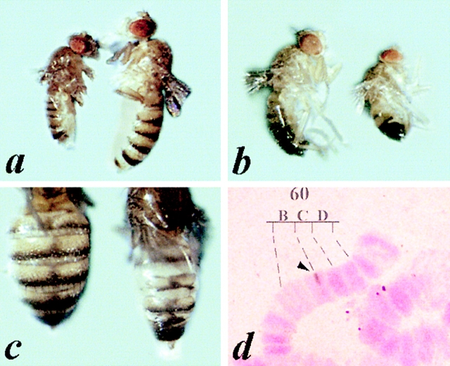
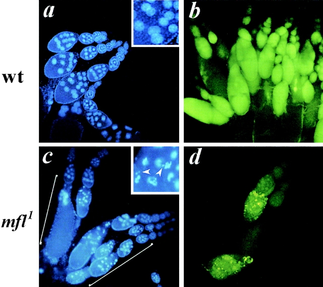
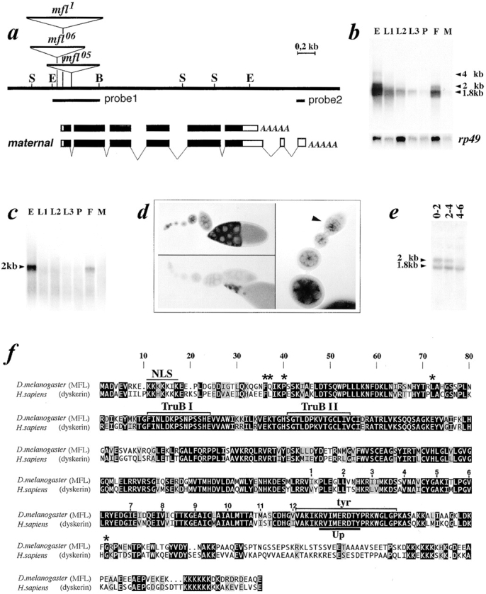
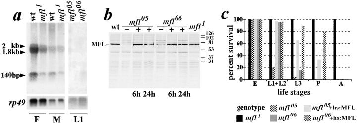
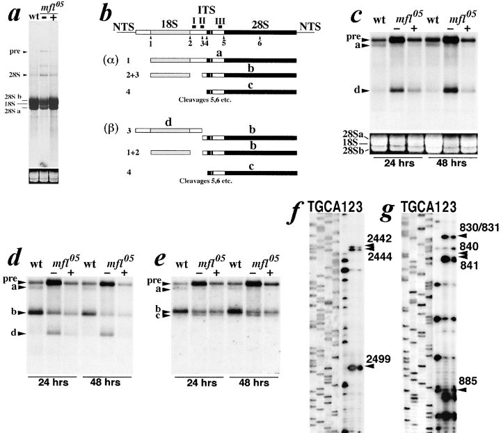
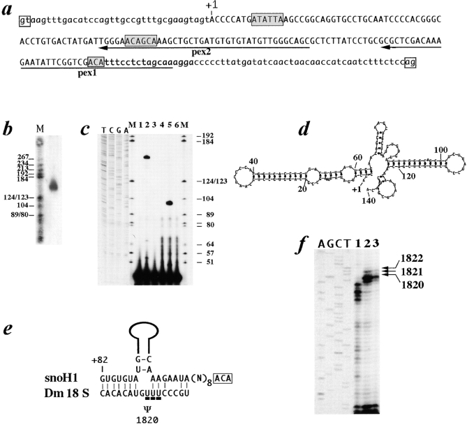
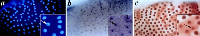
We report here the genetic, molecular, and functional characterization of the Drosophila melanogaster minifly (mfl) gene. Genetic analysis shows that mfl is essential for Drosophila viability and fertility. While P-element induced total loss-of-function mutations cause lethality, mfl partial loss-of-function mutations cause pleiotropic defects, such as extreme reduction of body size, developmental delay, hatched abdominal cuticle, and reduced female fertility. Morphological abnormalities characteristic of apoptosis are found in the ovaries, and a proportion of eggs laid by mfl mutant females degenerates during embryogenesis. We show that mfl encodes an ubiquitous nucleolar protein that plays a central role in ribosomal RNA processing and pseudouridylation, whose known eukaryotic homologues are yeast Cfb5p, rat NAP57 and human dyskerin, encoded by the gene responsible for the X-linked dyskeratosis congenita disease. mfl genetic analysis represents the first in vivo functional characterization of a member of this highly conserved gene family from higher eukaryotes. In addition, we report that mfl hosts an intron encoded box H/ACA snoRNA gene, the first member of this class of snoRNAs identified so far from Drosophila.


Figures

Figure 1
Figure 1
mfl1 phenotype. Comparing to wild-type, flies mfl1 females (a) and males (b) are both characterized by strong reduction of the body size, reduction in the number of abdominal bristles and abdominal cuticular defects; this last aspect is more marked in females (c). (d) Hybridization of a P-element probe to polytene chromosomes from mfl1 heterozygous larvae. The hybridization signal (arrowhead) is restricted to mfl1 parental chromosome of heterozygous larvae, allowing us to map the single P-element insertion at the 60B-60C polytene subdivisions boundary, on chromosome arm 2R.
Figure 2
Figure 2
Structure of mfl1 mutant ovaries. In the upper panel, as control, ovaries from wild-type females were stained with DAPI (a), or with the vital dye acridine orange (b). In the lower panel, ovaries from mfl1 homozygous females were stained with DAPI (c). Egg chambers, morphological abnormalities are observed beyond stage 7 of oogenesis (in brackets). Fragmented or condensed nurse cell nuclei with irregular shape are indicated by arrowheads in the boxed high magnification. (d) Acridine orange staining of mfl1 degenerating ovaries reveals highly fluorescent yellow spots, which correspond to apoptotic cells (Foley and Cooley, 1998).
Figure 3
Figure 3
Molecular characterization of the minifly gene. (a) Restriction map of the genomic region encompassing the minifly gene (S, SalI; E, EcoRI; B, BamHI). Genomic DNA sequence can be obtained from GenBank (accession number AF097634). On the top, position of P-element insertions. Below, organization of the 1.8- and 2.0-kb mfl transcription units. Exonic regions spanned by mfl ORF are depicted in black. Nucleotide sequence of the mfl maternal transcript can be obtained from GenBank (accession number AF089837). (b) Developmental Northern blot analysis of the mfl gene. Poly(A)+ RNA was hybridized to a genomic probe, depicted as probe 1 below the map of the region. E, 0–20 h embryos; L1, L2, and L3, first, second, and third instar larvae; P, pupae; F and M, female and male adult flies. The relative amount of RNA loaded in each lane was checked by hybridization with a probe derived from the gene coding for the Drosophila ribosomal protein rp49 (O'Connell and Rosbash, 1984). (c) Hybridization of the same RNA panel shown in b with probe 2 (depicted below the map), specific to mfl maternal mRNA. (d) In situ hybridization of whole mount wild-type ovaries with probe 2. On the left, hybridization with the mfl RNA anti-sense strand (top) and with the sense strand as negative control (bottom); on the right, enlargement of the tip of an ovariole in which the hybridization signal starts to be detected from the early oogenesis stages, within the germarial region (marked by the arrowhead). (e) Northern blot hybridization of total RNA preparations obtained from 0–2, 2–4, and 4–6 h staged embryos with genomic probe 1. (f) Alignment of MFL and dyskerin amino acid sequences. Black boxed letters highlight identical amino acids, different yet conserved amino acids are on a gray backgrounds; block letters on a white background indicate different and nonconserved amino acids. Lines above the sequences indicate putative functional domains; NLS, nuclear localization signal; TruBI and TruBII, regions having homology with bacterial and yeast tRNA pseudouridine synthases; tyr, tyrosine domain; Up, putative uracile binding pocket. Asterisks on the top indicate the positions of missense mutations so far identified in dyskerin from DKC patients (Heiss et al., 1998).
Figure 4
Figure 4
Molecular and functional characterization of mfl mutants. (a) Northern blot analysis of total RNA extracted from wild-type or mfl animals with a genomic probe including the fourth mfl intron. Female (F) and male (M) adult flies carrying the hypomorphic mfl1 allele or first-instar larvae (L1) carrying the mfl05 and mfl06 alleles were analyzed. (b) Western blot analysis of extracts obtained from wild-type or mfl animals, carrying (+) or not carrying (−) a MFL coding transgene. An affinity-purified rabbit polyclonal anti-MFL antibody, kindly provided by S. Poole, was used. Both wt and mfl homozygous animals were grown under heat shock regimen (30 min/d). As shown, MFL level is reduced in all mfl mutants (lanes −) but reaches nearly the wild-type amount in mfl05 and mfl06 transformed larvae (lanes +) at 6 or 24 h from the heat-shock pulse. (c) Lethal phase (see Materials and Methods) of mfl mutants is compared with that of mfl05 and mfl06 transgenic lines in which MFL was overexpressed from the heat-inducible hsp70 promoter. While most of mfl05 or mfl06 homozygotes develop only until the first or the third-larval instar, respectively, 30% of mfl05 and 80% of mfl06 transgenic animals reach the pupal stage when grown under daily heat-shock treatment. Moreover, these transgenic animals develop synchronously with their wild-type siblings and show a normal increase in their size.
Figure 5
Figure 5
(a) [3H]Uridine incorporation in wild-type or mfl05 larvae carrying (+) or not carrying (−) a MFL coding transgene under heat-shock treatment. Below, ethidium bromide staining of the 28Sa, 18S, and 28Sb rRNA species loaded in each lane. In Drosophila, 28S rRNA is cut to generate the 28Sa and 28Sb mature forms. (b) Genomic map of Drosophila rDNA. The two alternative α and β rRNA processing pathways (Long and Dawid, 1980) are depicted. In c–e, Northern analysis of total RNA from wild-type or mfl05 first instar larvae, at 24 or 48 h after egg hatching, with probes derived from the ITS region. The same blot was hybridized in c to probe I, in d to probe II, in e to probe III; probe positions are indicated by solid bars above the rDNA map. (f) Pseudouridylation of Drosophila 28S rRNA at positions U2442, U2444, U2499, and (g) of 18S rRNA, at positions U830/U831, U840, U841, U885. Wild-type untreated (lanes 1) and wild-type (lanes 2) or mfl05 (lanes 3) CMC-alkali treated RNAs were analyzed by primer extension using a 32P-labeled oligonucleotide complementary to the selected Drosophila rRNA sequences (see Materials and Methods). Lanes T, G, C, and A are dideoxy sequence reactions performed by using the same 28S or 18S primers on plasmids carrying the respective rDNA sequences.
Figure 6
Figure 6
Sequence, structure and properties of the mfl intron encoded snoH1 RNA. (a) Nucleotide sequence of the 235-bp-long fourth mfl intron; open boxes indicate the splicing sites. The estimated 5′ end of the snoH1 RNA, corresponding to position +37 of the mfl fourth intron, is indicated by +1. Shaded boxes indicate the two H and the 3′ terminal ACA element. SnoH1 nucleotide sequence can be obtained from GenBank (accession number AF089836). (b) Northern analysis of total RNA on a denaturing 6% polyacrylamide gel with a probe specific to mfl fourth intron (M, molecular weight marker V; Boehringer Mannheim). (c) 5′ end mapping of the snoH1 RNA by primer extension analysis of total larval RNA. The two oligonucleotides pex1 and pex2, complementary to the sequences underlined by the arrows in a, were used as primers. M, molecular weight marker V (Boehringer Mannheim). In lanes 1 and 4, no RNA was loaded as internal control. In lanes 2 and 3, the RNA was incubated with oligonucleotide pex1, with or without reverse transcriptase. In lanes 5 and 6, the RNA was incubated with oligonucleotide pex2, with or without reverse transcriptase. (d) Predicted secondary structure of the mfl intron encoded RNA. (e) Potential base-pairing interactions between the snoH1 RNA and Drosophila 18S rRNA sequences. The upper strand represents the snoH1 RNA sequence in a 5′ to 3′ orientation. Solid lines schematically represent the hairpin domain of the snoH1 RNA. The 3′ terminal ACA motif is boxed. The ribosomal pseudouridine potentially selected by the snoH1 RNA is indicated by Ψ. (f) Pseudouridylation of Drosophila 18S rRNA, at positions U1820, U1821, and U1822. Untreated (lane 1) or CMC-alkali treated (lanes 2 and 3) RNAs were analyzed by primer extension using an oligonucleotide complementary to the selected Drosophila 18S rRNA sequences (see Materials and Methods). In lanes 1 and 2, RNA was extracted from wild-type larvae; on lane 3, from mfl05 larvae. Lanes A, G, C, and T are dideoxy sequence reactions performed by using the same primer and a plasmid carrying Drosophila 18S rRNA sequences.
Figure 7
Figure 7
Intracellular distribution of snoH1 RNA and MFL protein in Drosophila ovaries. (a) Nuclei were counterstained with DAPI. (b) In situ hybridization of a snoH1 RNA antisense probe exclusively labeled the nucleoli. (c) Immunohistochemical localization of MFL protein in wild-type ovaries with an affinity-purified rabbit polyclonal anti-MFL antibody (kindly provided by S. Poole). The protein shows a specific nucleolar localization, although occasional intracytoplasmatic diffusion can be observed.

References

    1. Abrams JM, White K, Fessler LI, Steller H. Programmed cell death during Drosophilaoogenesis. Development. 1993;117:29–43. - PubMed
    1. Ashburner, M. 1989. Drosophila: A Laboratory Manual. Cold Spring Harbor, NY, Cold Spring Harbor Laboratory Press. 434 pp.
    1. Bachellerie JP, Cavaille J. Guiding ribose methylation of rRNA. Trends Biochem Sci. 1997;22:257–261. - PubMed
    1. Bakin A, Ofengand J. Four newly located pseudouridylate residues in Escherichia coli23S ribosomal RNA are all at the peptidyltransferase center: analysis by the application of a new sequencing technique. Biochemistry. 1993;32:9754–9762. - PubMed
    1. Balakin AG, Smith L, Fournier MJ. The RNA world of the nucleolus: two major families of small RNAs defined by different box elements with related functions. Cell. 1996;86:823–834. - PubMed

Publication types

MeSH terms

Associated data