. 1999 Mar 22;144(6):1163-72.
doi: 10.1083/jcb.144.6.1163.

Recombinant major vault protein is targeted to neuritic tips of PC12 cells

Affiliations

Recombinant major vault protein is targeted to neuritic tips of PC12 cells

C Herrmann et al. J Cell Biol. .

Abstract

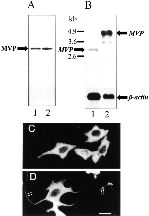
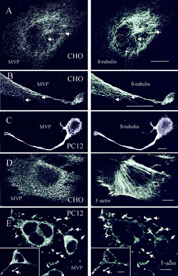
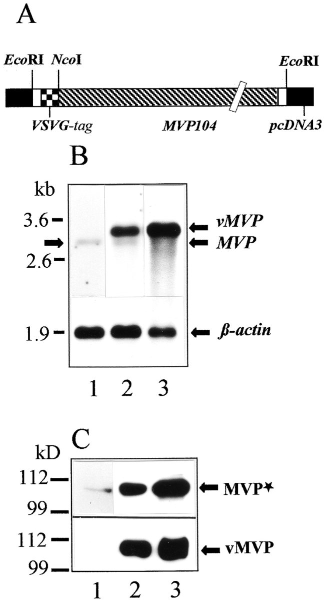
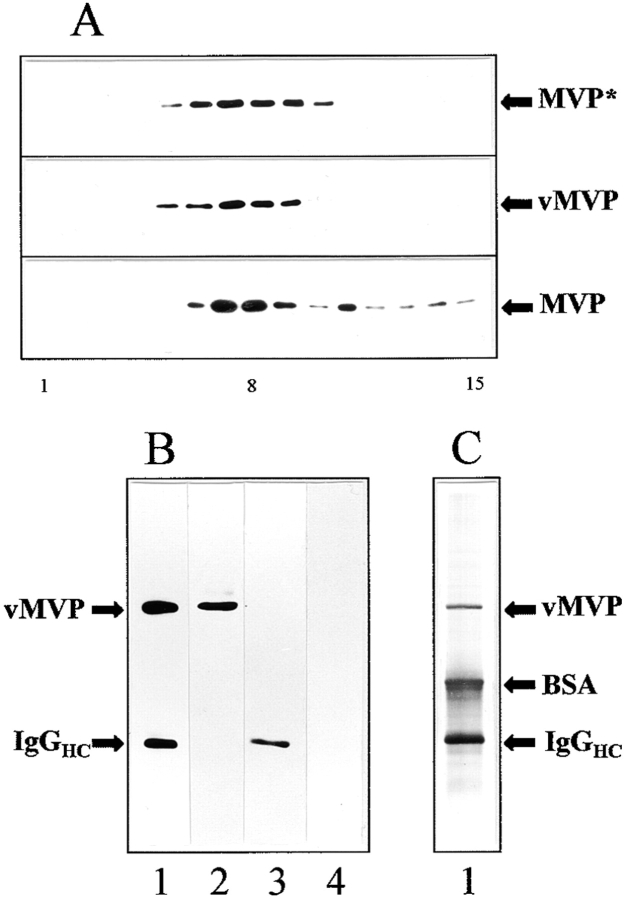
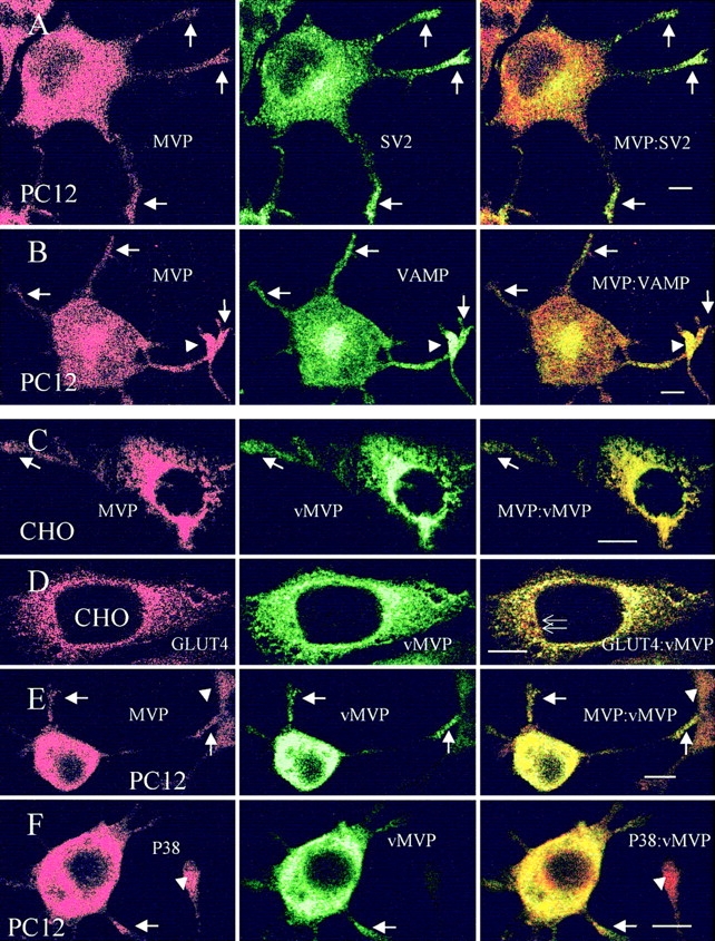
The major vault protein (MVP) is the predominant constituent of ubiquitous, evolutionarily conserved large cytoplasmic ribonucleoprotein particles of unknown function. Vaults are multimeric protein complexes with several copies of an untranslated RNA. Double labeling employing laser-assisted confocal microscopy and indirect immunofluorescence demonstrates partial colocalization of vaults with cytoskeletal elements in Chinese hamster ovary (CHO) and nerve growth factor (NGF)-treated neuronlike PC12 cells. Transfection of CHO and PC12 cells with a cDNA encoding the rat major vault protein containing a vesicular stomatitis virus glycoprotein epitope tag demonstrates that the recombinant protein is sorted into vault particles and targeted like endogenous MVPs. In neuritic extensions of differentiated PC12 cells, there is an almost complete overlap of the distribution of microtubules and vaults. A pronounced colocalization of vaults with filamentous actin can be seen in the tips of neurites. Moreover, in NGF-treated PC12 cells the location of vaults partially coincides with vesicular markers. Within the terminal tips of neurites vaults are located near secretory organelles. Our observations suggest that the vault particles are transported along cytoskeletal-based cellular tracks.


Figures

Figure 1
Figure 1
Expression of MVP in CHO and PC12 cells. (A) Western blot of total protein extracts of CHO (lane 1) and PC12 (lane 2) cells using polyclonal affinity-purified anti–rat vault antibody. 50 μg of protein was loaded per lane. (B) Northern blot of mRNA isolated from CHO (lane 1) and PC12 (lane 2) cells using a DIG-labeled RNA encoding rat MVP as a probe. 800 ng of mRNA was applied per lane. The hybridization signal for β-actin (lower graph) served as a control. Sizes of RNA markers are indicated (left). Immunocytochemistry of CHO (C) and PC12 (D) cells using the same antibody as in B demonstrated the overall distribution of vault particles. In D, localization of vaults in neuritic extensions and tips is indicated (arrows). Bar, 20 μm.
Figure 2
Figure 2
Distribution of vaults and β-tubulin or F-actin in CHO and PC12 cells. The localization was analyzed by double labeling and confocal microscopy of median sections using polyclonal affinity-purified anti–rat vault antibody, anti–β-tubulin mAb, or TRITC-conjugated phalloidin decorating filamentous actin. (A and B) Arrows indicate colocalization of vault particles and microtubules in CHO cells. (C) Overlapping distribution of vaults and microtubules in differentiated PC12 cells is shown. (D) Localization of vaults and filamentous actin in CHO cells. (E) Distribution of vaults and F-actin in PC12 cells is shown. Only in neuritic tips distribution of vaults and F-actin overlap (arrows and inset). Bars, 10 μm.
Figure 3
Figure 3
Transient expression of rat vMVP in CHO cells. (A) Schematic drawing of the engineered construct denoted pvMVP for heterologous expression of rat MVP in mammalian cell lines is shown. Note that expression vector and cDNA sequence encoding MVP are not in scale. (B) Northern blot using a DIG-labeled RNA encoding rat MVP as a probe is shown. 800 ng of mRNA was applied per lane. Lane 1 shows mRNA isolated from nontransfected CHO cells; lane 2 shows mRNA from CHO cells transfected with pvMVP; and lane 3 shows mRNA from transfected CHO cells treated with sodium butyrate. The hybridization signal for β-actin (lower graph) served as a control. Arrows indicate the position of the endogenous MVP transcript (left and right), overexpressed vMVP, and β-actin (both right). The position of marker RNA is given in kilobases. (C) Western blot using total protein extract of CHO cells, polyclonal affinity-purified anti–rat vault antibody (upper graph), or anti-VSVG mAb (lower graph) is shown. (Lane 1) Nontransfected; (lane 2) pvMVP-transfected, and (lane 3) pvMVP-transfected and butyrate-treated cells. 30 μg of protein was applied per lane. Protein bands detected by anti-MVP antibody (MVP*) or anti-VSVG antibody (vMVP) are marked by arrows (right). Note, the anti-MVP antibody detects endogenous and recombinant MVP. The size of marker polypeptides is given in kilodaltons (left).
Figure 4
Figure 4
Recombinant MVP is assembled into vault particles: subcellular fractionation (A) and immunoprecipitation of VSVG-tagged major vault protein (B and C). (A) Western blot analysis of the sedimentation behavior of vaults derived from CHO cells on glycerol gradients is shown. The following are shown: (top) Immunodetection of endogenous and recombinant MVP (MVP*) of pvMVP-transfected CHO cells using the anti–rat vault antibody; (middle) immunodetection of vMVP using the anti-VSVG antibody. In the top and middle frames identical fractions were applied per lane. The bottom frame shows immunodetection of endogenous MVP derived from nontransfected cells using the anti–rat vault antibody. Note that both the amount of material loaded per lane as well as the exposure time was doubled in the bottom frame blot as compared to the top two (50:100 μg protein per gradient, 2:4 min exposure time). Fractions of 300 μl were collected from the gradient and analyzed by immunoblotting. The number of fraction is indicated below the graph: starting fraction on top of the gradient (1) vault-enriched fraction (8) and bottom fraction (15). (B) Western blot and (C) silver staining of immunoprecipitate using anti-VSVG mAb and anti–mouse IgG-coated magnetic beads as a solid carrier (lane 1). Bands comprising minor vault proteins are not visible. Parent fraction of transfected cells before immunoisolation (lane 2). Immunoprecipitate of nontransfected CHO cells applying an identical experimental procedure as for transfected cells (lane 3). Parent fraction of nontransfected cells before immunoisolation (lane 4). Positions of vMVP, BSA, and heavy chains of mouse immunoglobulins (IgGHC) are indicated.
Figure 5
Figure 5
Localization of vault particles and vesicle markers in CHO and PC12 cells. Distribution of endogenous vaults and the synaptic vesicle marker SV2 (A) and VAMP/synaptobrevin (B) in differentiated PC12 cells is shown by double labeling in confocal images of middle sections. Arrows indicate colocalization of secretory organelles and vaults in neuritic tips. Arrowheads indicate immunostained neuritic tip of adjacent cell. An identical distribution of endogenous and recombinant MVP (vMVP) is observed in CHO (C) and differentiated PC12 (E) cells. Note, that antivault antibody (MVP) recognizes both endogenous and recombinant MVP, whereas anti-VSVG mAb only detects vMVP (C and E). Arrows indicate targeting of endogenous and recombinant MVP to cell extensions. Arrowheads indicate processes of neighboring cells revealing no or low expression of vMVP. (D) Localization of vMVP as compared to GLUT4-containing organelles is shown. Thin arrows indicate the differential distribution of GLUT4 and MVP in the perinuclear region of CHO cells. (F) Colocalization of the synaptic vesicle marker P38/synaptophysin and vMVP in neuritic extensions of differentiated PC12 cells (arrows). Arrowheads show the nontransfected cells. Bars, 10 μm.

References

    1. Abbondanza C, Rossi V, Roscigno A, Gallo L, Belsito A, Piluso G, Medici N, Nigro V, Molinari AM, Moncharmont B, Puca GA. Interaction of vault particles with estrogen receptor in the MCF-7 breast cancer cell. J Cell Biol. 1998;141:1301–1310. - PMC - PubMed
    1. Bassel GJ, Powers CM, Taneja KL, Singer RH. Single mRNAs visualized by ultrastructural in situ hybridization are principally localized at actin filament intersections in fibroblasts. J Cell Biol. 1994a;126:863–876. - PMC - PubMed
    1. Bassel GJ, Singer RH, Kosik KS. Association of poly(A+) mRNA with microtubules in cultured neurons. Neuron. 1994b;12:571–582. - PubMed
    1. Carson JH, Worboys K, Ainger K, Barbarese E. Translocation of myelin basic protein mRNA in oligodendrocytes requires microtubules and kinesin. Cell Motil Cytoskel. 1997;38:318–328. - PubMed
    1. Chugani DC, Rome LH, Kedersha NL. Evidence that vault ribonucleoprotein particles localize to the nuclear pore complex. J Cell Sci. 1993;106:23–29. - PubMed

Publication types