. 1999 Mar 22;144(6):1235-44.
doi: 10.1083/jcb.144.6.1235.

Rho GTPases control polarity, protrusion, and adhesion during cell movement

Affiliations

Rho GTPases control polarity, protrusion, and adhesion during cell movement

C D Nobes et al. J Cell Biol. .

Abstract

Cell movement is essential during embryogenesis to establish tissue patterns and to drive morphogenetic pathways and in the adult for tissue repair and to direct cells to sites of infection. Animal cells move by crawling and the driving force is derived primarily from the coordinated assembly and disassembly of actin filaments. The small GTPases, Rho, Rac, and Cdc42, regulate the organization of actin filaments and we have analyzed their contributions to the movement of primary embryo fibroblasts in an in vitro wound healing assay. Rac is essential for the protrusion of lamellipodia and for forward movement. Cdc42 is required to maintain cell polarity, which includes the localization of lamellipodial activity to the leading edge and the reorientation of the Golgi apparatus in the direction of movement. Rho is required to maintain cell adhesion during movement, but stress fibers and focal adhesions are not required. Finally, Ras regulates focal adhesion and stress fiber turnover and this is essential for cell movement. We conclude that the signal transduction pathways controlled by the four small GTPases, Rho, Rac, Cdc42, and Ras, cooperate to promote cell movement.


Figures
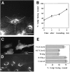
Figure 5
Figure 5
Cdc42 is required for reorientation of the Golgi apparatus. (A) REFs fixed and stained using indirect immunofluorescence to show the distribution of the Golgi apparatus 4 h after wounding. The percentage of cells having their Golgi apparatus in the forward-facing 120° sector was measured 2, 4, and 6 h after wounding (B). Random orientation of the Golgi apparatus with respect to the wound edge corresponds to 33% at 0 h (B). Wound edge cells were microinjected with expression vectors encoding myc-tagged V12Cdc42, N17Cdc42, WASp fragment, or N17Rac 1 h after wounding. Expression of myc-tagged proteins could first be detected 1 h later, at which time 49% of first row control wound edge cells had their Golgi apparatus facing the wound. Cells were fixed 6 h after wounding and double-labeled to reveal expression of myc-tagged proteins and the Golgi apparatus (shown for myc-tagged WASp fragment in C and corresponding Golgi apparatus in D). Percentage of cells with Golgi apparatus facing the wound was calculated for each condition (E). For each time point and/ or experimental condition, at least 100 cells from three different experiments were scored. Values represent means ± SEM. Bar, 20 μm.
Figure 1
Figure 1
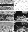
REF cell behavior at the wound edge. REF monolayers were fixed 5 min (A) and 5 h (B) after wounding and visualized with rhodamine-conjugated phalloidin. Bar, 120 μm. (C) Phase-contrast image of wound edge 3 h after wounding showing polarized morphology of front row cells. Bar, 20 μm. (D) Outline of three cells drawn from time-lapse recording after 15 min (dashed), 2 h, and 4 h as they migrate into the wound space (see numbers 1, 2, and 4 within nucleus of cell on left side). Dashed line across wound space is 125 μm. (E and F) High-power image of wound edge cell fixed 3 h after wounding and costained for actin filaments (E) and for phosphotyrosine (F). Wound edge cells display actin stress fibers, lamellipodia, filopodia (generally within lamellipodia), elongated focal adhesions, and focal complexes (arrow in F). Bar, 25 μm.
Figure 2
Figure 2
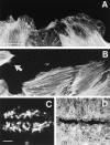
Rac is required for lamellipodial protrusions. Control wounds (A) and N17Rac (1 mg/ml)-injected wounds (B–D) were fixed and actin filaments visualized with rhodamine-conjugated phalloidin. Arrow in B indicates filopodia extending from an N17Rac-injected cell. Bar in A, 20 μm. Wound assay from which measurements of wound closure are made is shown in C and D. 1 h after wounding, wound edge cells were coinjected with N17Rac and injection marker (fluorescent dextran, C). Wounds were fixed ∼5–6 h later when the control injected wounds had closed. Bar in C, 200 μm.
Figure 3
Figure 3
Effects of activating or inhibiting Rac and Cdc42 on wound closure. REF monolayers were wounded and the activity of Rac and Cdc42 in wound edge cells was modulated by microinjection of N17Rac (1 mg/ml), N17Cdc42 (2 mg/ml), WASp fragment (2 mg/ml), V12Rac (1 mg/ml), V12Cdc42 (1 mg/ml) proteins, or expression vectors encoding the same (indicated by DNA in parentheses). Percent wound closure was calculated as described in Materials and Methods. Experimental wounds were fixed at the same time that control wounds closed and stained with rhodamine-conjugated phalloidin to visualize actin filaments and wound space. Cells were coinjected with fluorescent dextran in order to visualize the position of injected wound edge cells. Values represent means ± SEM for at least three independent experiments.
Figure 4
Figure 4
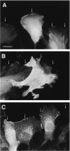
Cdc42 is required for morphological polarization in leading edge cells. 1 h after wounding, wound edge REFs were microinjected with fluorescent dextran (2 mg/ml) (A) or a myc-tagged expression vector encoding WASp fragment to inhibit Cdc42 activity (B) or a myc-tagged expression vector encoding V12Cdc42 (C). Cells were fixed 3 h later when wounds were still open and myc-tagged proteins were visualized by indirect immunofluorescence. The position of the wound edge in A–C is indicated by arrows. Bar, 20 μm.
Figure 6
Figure 6
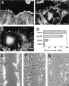
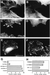
Rho is required for cell substrate adhesion but focal adhesions and stress fibers inhibit movement. Wounded REFs untreated (A) or injected with C3 transferase (0.07 mg/ml) 1 h after wounding (B and C) were fixed 2 h later and actin filaments were visualized with rhodamine-conjugated phalloidin (A and B) and vinculin was visualized by indirect immunofluorescence (C). (D) Percent wound closure was calculated for wounds injected with dextran (Control), C3 transferase (0.07 and 0.3 mg/ml), and V14Rho protein (0.4 mg/ml). Values represent means ± SEM for at least three different experiments. Bar, 20 μm. Phase-contrast images of wounded REFs 15 min after wounding (E) and 4 h after wounding in the presence (F) or absence (G) of Y-27632 (20 μm). Bar, 100 μm.
Figure 7
Figure 7
Ras and ERK are activated after wounding. REF wounds were fixed 40 s, 5, 10, 20, and 60 min after wounding. Dually phosphorylated ERK was visualized by indirect immunofluorescence. Cells were pretreated with the MEK inhibitor PD98059 (60 μM) before wounding and then fixed 10 min after wounding and stained to reveal phosphoERK (10′+PD). Bar, 100 μm.
Figure 8
Figure 8
Ras is required for cell movement. 1 h after wounding, wound edge cells were coinjected with the neutralizing Ras antibody Y13-259 (9 mg/ml) and fluorescent dextran as an injection marker (A–D). Cells were fixed 3–4 h later and actin filaments were visualized with rhodamine-conjugated phalloidin (A) and vinculin was visualized by indirect immunofluorescence (C). Arrows in C indicate larger focal adhesions in the Ras antibody– injected cell. Injection marker is shown in B and D. Expressed Ras was localized using indirect immunofluorescence (E) and cells were costained to reveal vinculin (F). Percent wound closure was calculated for wounds injected with V14Rho (0.4 mg/ml), anti-Ras antibody (8 mg/ml), anti-Ras antibody (8 mg/ml) + C3 transferase (0.06 mg/ml), anti-Ras antibody (8 mg/ml) in the presence of Y-27632 (20 μm) (Anti-Ras+Y), and dextran (Control) (G). Wounds were pretreated with inhibitors PD98059 (60 μM) and LY294002 (20 μM) for 20 min before wounding. Percent wound closure for these inhibitors compared with control wounds is shown in H. Bar, 20 μm.

References

    1. Adams AE, Johnson DI, Longnecker RM, Sloat BF, Pringle JR. CDC42 and CDC43, two additional genes involved in budding and establishment of cell polarity in the yeast Saccharomyces cerevisiae. . J Cell Biol. 1990;111:131–142. - PMC - PubMed
    1. Allen WE, Zicha D, Ridley AJ, Jones GE. A role for Cdc42 in macrophage chemotaxis. J Cell Biol. 1998;141:1147–1157. - PMC - PubMed
    1. Amano M, Chihara K, Kimura K, Fukata Y, Nakamura N, Mastuura Y, Kaibuchi K. Formation of actin stress fibers and focal adhesions enhanced by Rho-kinase. Science. 1997;275:1308–1311. - PubMed
    1. Anand-Apte B, Zetter BR, Viswanathan A, Qui R, Chen J, Ruggieri R, Symons M. Platelet-derived growth factor and fibronectin-stimulated migration are differentially regulated by the Rac and extracellular signal-regulated kinase pathways. J Biol Chem. 1997;272:30688–30692. - PubMed
    1. Bershadsky AD, Futerman AH. Disruption of the Golgi apparatus by brefeldin A blocks cell polarization and inhibits directed cell migration. Proc Natl Acad Sci USA. 1994;91:5686–5689. - PMC - PubMed

Publication types