. 1999 Mar 22;144(6):1245-58.
doi: 10.1083/jcb.144.6.1245.

Role of proteins of the Ena/VASP family in actin-based motility of Listeria monocytogenes

Affiliations

Role of proteins of the Ena/VASP family in actin-based motility of Listeria monocytogenes

V Laurent et al. J Cell Biol. .

Abstract

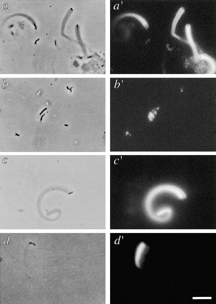
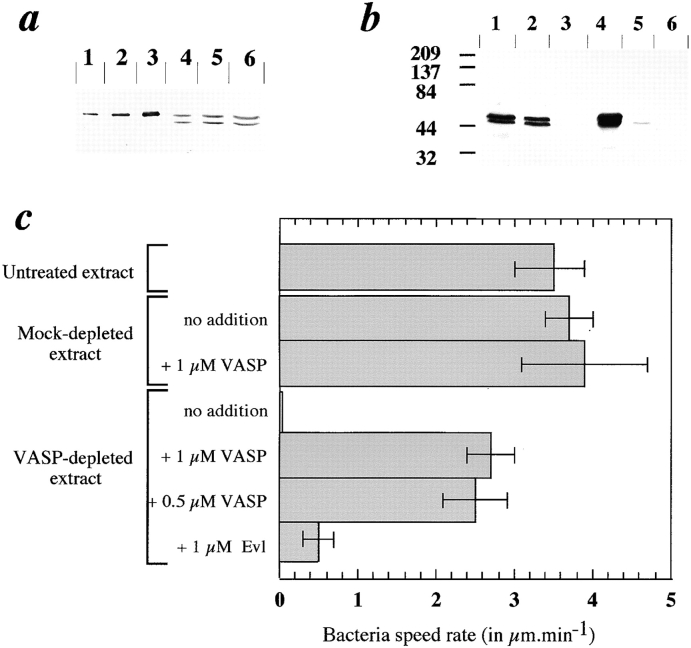
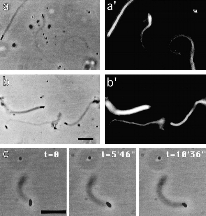
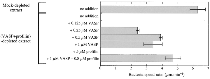
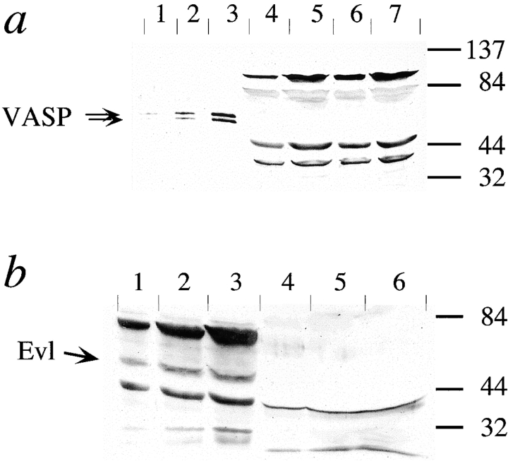
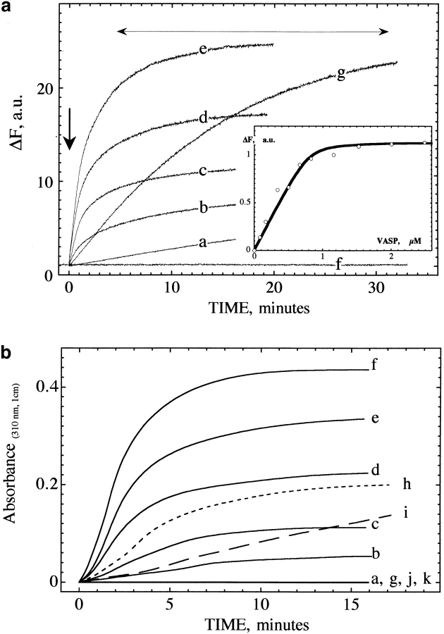
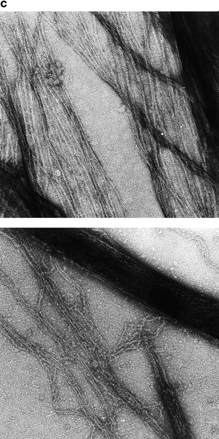
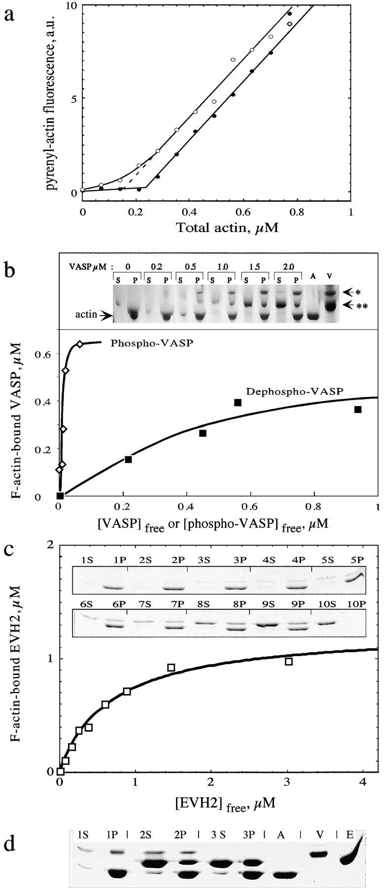
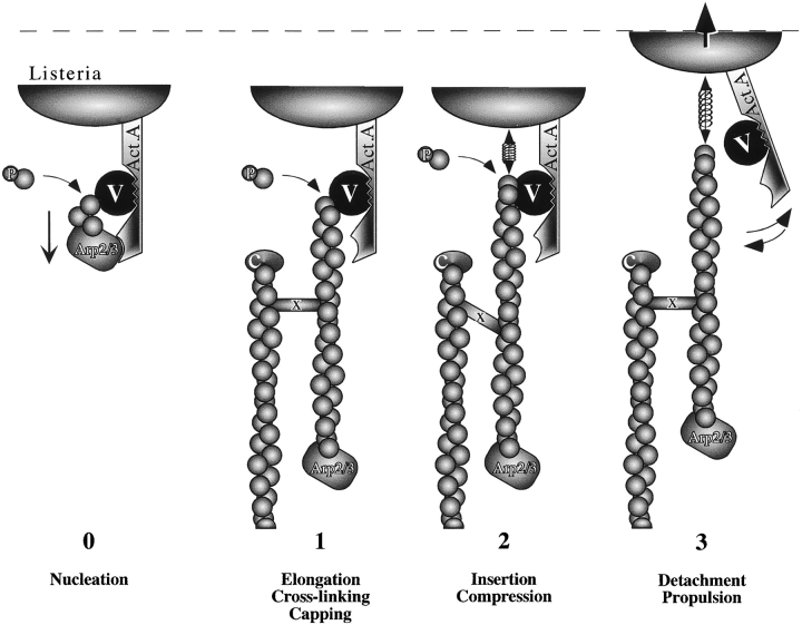
Intracellular propulsion of Listeria monocytogenes is the best understood form of motility dependent on actin polymerization. We have used in vitro motility assays of Listeria in platelet and brain extracts to elucidate the function of the focal adhesion proteins of the Ena (Drosophila Enabled)/VASP (vasodilator-stimulated phosphoprotein) family in actin-based motility. Immunodepletion of VASP from platelet extracts and of Evl (Ena/VASP-like protein) from brain extracts of Mena knockout (-/-) mice combined with add-back of recombinant (bacterial or eukaryotic) VASP and Evl show that VASP, Mena, and Evl play interchangeable roles and are required to transform actin polymerization into active movement and propulsive force. The EVH1 (Ena/VASP homology 1) domain of VASP is in slow association-dissociation equilibrium high-affinity binding to the zyxin-homologous, proline-rich region of ActA. VASP also interacts with F-actin via its COOH-terminal EVH2 domain. Hence VASP/ Ena/Evl link the bacterium to the actin tail, which is required for movement. The affinity of VASP for F-actin is controlled by phosphorylation of serine 157 by cAMP-dependent protein kinase. Phospho-VASP binds with high affinity (0.5 x 10(8) M-1); dephospho-VASP binds 40-fold less tightly. We propose a molecular ratchet model for insertional polymerization of actin, within which frequent attachment-detachment of VASP to F-actin allows its sliding along the growing filament.


Figures

Figure 1
Figure 1
VASP is essential for actin-based motility of Listeria in platelet extracts. Actin structures formed by L. monocytogenes in: a and a′, mock-depleted platelet extract; b and b′, VASP-depleted extract (two cycles of depletion on anti-VASP–coated Dynabeads); c and c′, VASP-depleted extract supplemented with 1 μM bacterial recombinant VASP; d and d′, VASP-depleted extract supplemented with 0.9 μM eukaryotic recombinant VASP. (a–d) Phase-contrast; (a′–d′) corresponding rhodamine-actin fluorescence. Bar, 5 μm.
Figure 2
Figure 2
Immunodepletion of VASP from platelet extracts and Listeria movement in VASP-depleted extracts supplemented with recombinant VASP or Evl. (a) Quantitation of VASP in platelet extracts. 0.5 (lane 1), 1 (lane 2), and 2 pmol (lane 3) of recombinant VASP and 0.66 (lane 4), 1.33 (lane 5), and 2.66 μl (lane 6) of platelet extract were electrophoresed and immunoblotted using anti-VASP (M4 polyclonal antibody). Comparison of the densitometric patterns of the standards and of platelet VASP using the NIH Image analysis software led to an estimate of 0.7 μM VASP in the extracts. (b) Analysis of the immunodepletion of VASP from platelet extracts. Lane 1, whole extract; lane 2, mock-depleted extract; lane 3, VASP-depleted extract (two cycles of depletion using Dynabeads); lane 4, Dynabead-bound VASP after the first cycle of depletion; lane 5, Dynabead-bound VASP after the second cycle of depletion; and lane 6, control beads (not coated with anti-VASP) used for the mock depletion. The Western blot was revealed using M4 polyclonal anti-VASP. (c) Mean rates of movement of Listeria in VASP-depleted extracts and in depleted extracts supplemented with VASP or Evl recombinant proteins. 10–15 rate measurements were performed for each sample. At least three independent experiments were carried out.
Figure 3
Figure 3
Movement of L. monocytogenes in mouse brain extracts from wild-type and Mena (−/−) animals. Typical actin tails formed in wild-type (a and a′) and in Mena (−/−) (b and b′) mouse brain extracts. (a and b) Phase-contrast; (a′ and b′) rhodamine-actin fluorescence. c shows a time-lapse illustration of the movement. Bar, 5 μm.
Figure 4
Figure 4
Evl is essential for actin-based motility of Listeria in mouse brain extracts. (a) Western blot analysis of Evl depletion from Mena (−/−) mouse brain extracts. Lane 1, 15 pmol of recombinant Evl; lane 2, 10 μl mock-depleted extract; lane 3, 10 μl Evl-depleted extract; lane 4, Dynabead-bound Evl after the first cycle of depletion; and lane 5, control Dynabeads used for the mock depletion. The blot was revealed using an anti-Evl mAb. (b) Effect of Evl depletion on Listeria movement in wild-type brain extracts. The percentages of motile bacteria, defined as: (number of motile bacteria/number of total bacteria) × 100 (dark gray bars), and of immotile bacteria that were surrounded by actin clouds, defined as: (number of nonmotile bacteria surrounded by actin clouds/number of total bacteria) × 100 (light gray bars), were determined under each condition. 271 ± 24 bacteria were counted for each measurement. Motile bacteria moved at rates that fell in the range of 0.16 ± 0.06 μm/min in all samples. (c) Effect of Evl depletion on Listeria movement in Mena (−/−) mouse brain extracts. Data are represented as in b. 282 ± 49 bacteria were counted for each measurement.
Figure 5
Figure 5
Profilin is not essential for actin-based motility of Listeria in platelet extracts. The mean rate of movement of Listeria was measured in mock-depleted extracts, profilin-depleted extracts, profilin + VASP double- depleted extracts, supplemented with the indicated amounts of recombinant VASP or purified bovine profilin. Mean rate values result from the average of 10 measurements.
Figure 6
Figure 6
Immunodetection of VASP in mouse brain extracts and of Evl in platelet extracts. (a) Platelet extract (1, 2, and 4 μl, corresponding to 0.7, 1.4, and 2.1 pmol of VASP, in lanes 1, 2, and 3, respectively), Mena (−/−) mouse brain extracts (10 and 20 μl in lanes 4 and 5), and wild-type mouse brain extracts (10 and 20 μl in lanes 6 and 7) were analyzed by immunoblotting using polyclonal M4 anti-VASP antibody. Assuming that the limit of detection is 0.4 pmol, the data indicate that the maximum concentration of VASP in brain extracts is 20 nM, a concentration too low to support Listeria movement. (b) Wild-type mouse brain extracts (5, 10, and 20 μl corresponding to 3.5, 7, and 14 pmol of Evl in lanes 1, 2, and 3, respectively) and 10, 20, and 40 μl platelet extracts (lanes 4, 5, and 6, respectively) were analyzed by immunoblotting using a polyclonal anti-Evl antibody. Assuming that the lower limit of detection is 2 pmol Evl, the data indicate that the maximum concentration of Evl in platelets is 50 nM, which is too low to support motility of Listeria.
Figure 7
Figure 7
VASP induces polymerization of G-actin into F-actin bundles. (a) Pyrenyl-actin fluorescence measurements. 1 μM pyrenyl-labeled MgATP-G-actin was supplemented, at time indicated by the vertical arrow, with eukaryotic recombinant VASP at the following concentrations (μM): a, 0; b, 0.25; c, 0.5; d, 1; e, 2; f, 1 μM VASP + 0.1 M KCl; g, 0 VASP, 2 μM GST-EVH2. The fluorescence of 1 μM pyrenyl-G-actin is equal to 1 by convention. The horizontal arrow indicates the fluorescence measured for 1 μM F-actin polymerized in standard F buffer (0.1 M KCl, 1 mM MgCl2 added to G buffer). (Inset) Extent of fluorescence change at the end of the polymerization process of 0.7 μM MgATP-G-actin induced by VASP at the indicated concentrations. Fluorescence was measured 1 h after the preparation of the samples. Final conditions were: 5 mM Tris-Cl, pH 7.5, 0.2 mM ATP, 0.1 mM CaCl2, 1 mM DTT, 0.2 mM EGTA, 50 μM MgCl2, 2 mM Hepes, 15 mM NaCl, 1% glycerol, 20°C. (b) Turbidity measurements at 310 nm. 1 μM MgATP-G-actin (same actin solution as in a) was supplemented, at time 0, with eukaryotic recombinant VASP at the following concentrations (μM): a, 0; b, 0.29; c, 0.58; d, 1.16; e, 1.74; f, 2.34; g, 0 VASP, 2 μM GST-EVH2; h, 1.16 μM VASP + 2 μM GST-EVH2; i, 1.16 μM VASP + 0.05 M KCl; j, 1.16 μM VASP + 0.1 M KCl; k, no actin, 1.16 μM VASP. Ionic conditions were as in a. Temperature was 20°C. Optical path length = 1 cm. (c) Actin bundles assembled from Mg-actin in the presence of VASP. Samples of MgATP-G-actin (1 μM) preincubated for 10 min with 1 μM VASP were negatively stained and observed in the electron microscope. (Top) Bacterial recombinant VASP. (Bottom) Eukaryotic recombinant VASP.
Figure 7
Figure 7
VASP induces polymerization of G-actin into F-actin bundles. (a) Pyrenyl-actin fluorescence measurements. 1 μM pyrenyl-labeled MgATP-G-actin was supplemented, at time indicated by the vertical arrow, with eukaryotic recombinant VASP at the following concentrations (μM): a, 0; b, 0.25; c, 0.5; d, 1; e, 2; f, 1 μM VASP + 0.1 M KCl; g, 0 VASP, 2 μM GST-EVH2. The fluorescence of 1 μM pyrenyl-G-actin is equal to 1 by convention. The horizontal arrow indicates the fluorescence measured for 1 μM F-actin polymerized in standard F buffer (0.1 M KCl, 1 mM MgCl2 added to G buffer). (Inset) Extent of fluorescence change at the end of the polymerization process of 0.7 μM MgATP-G-actin induced by VASP at the indicated concentrations. Fluorescence was measured 1 h after the preparation of the samples. Final conditions were: 5 mM Tris-Cl, pH 7.5, 0.2 mM ATP, 0.1 mM CaCl2, 1 mM DTT, 0.2 mM EGTA, 50 μM MgCl2, 2 mM Hepes, 15 mM NaCl, 1% glycerol, 20°C. (b) Turbidity measurements at 310 nm. 1 μM MgATP-G-actin (same actin solution as in a) was supplemented, at time 0, with eukaryotic recombinant VASP at the following concentrations (μM): a, 0; b, 0.29; c, 0.58; d, 1.16; e, 1.74; f, 2.34; g, 0 VASP, 2 μM GST-EVH2; h, 1.16 μM VASP + 2 μM GST-EVH2; i, 1.16 μM VASP + 0.05 M KCl; j, 1.16 μM VASP + 0.1 M KCl; k, no actin, 1.16 μM VASP. Ionic conditions were as in a. Temperature was 20°C. Optical path length = 1 cm. (c) Actin bundles assembled from Mg-actin in the presence of VASP. Samples of MgATP-G-actin (1 μM) preincubated for 10 min with 1 μM VASP were negatively stained and observed in the electron microscope. (Top) Bacterial recombinant VASP. (Bottom) Eukaryotic recombinant VASP.
Figure 8
Figure 8
VASP binds to and stabilizes F-actin in a phosphorylation-regulated manner. (a) VASP shifts the critical concentration plots for actin assembly toward a lower value. Critical concentration plots for polymerization of Mg-actin in the absence (filled circles) and presence (open circles) of 1 μM eukaryotic recombinant VASP. Pyrenyl-labeled actin was polymerized at a high concentration and serially diluted in buffer adjusted to the following ionic conditions: 5 mM Tris-Cl, pH 7.5, 0.2 mM ATP, 0.1 mM CaCl2, 0.2 mM EGTA, 0.1 mM MgCl2, 1 mM DTT, 2 mM Hepes, 15 mM NaCl, and 1% glycerol. (b) Sedimentation assay for the binding of eukaryotic recombinant VASP to F-actin. Mg-F-actin was polymerized under physiological ionic conditions (0.1 M KCl, 1 mM MgCl2) and mixed at 1 μM with VASP at the indicated concentrations. Samples were centrifuged for 30 min at 400,000 g, 20°C. Pellets were resuspended in G buffer at the original volume. Pellets (P) and supernatants (S) were submitted to SDS-PAGE. A, actin alone (1 μM); V, VASP alone (2 μM); *, 50-kD band corresponding to serine 157–phosphorylated VASP; **, 46-kD band corresponding to serine 157–unphosphorylated VASP. Data are plotted as bound VASP versus free VASP. The concentrations of free and F-actin–bound VASPs were derived from the scanning of the gels (see Materials and Methods). The curves represent the fit to the data using values of equilibrium dissociation constants K Y = 22 nM and K X = 0.8 μM for phospho- and dephospho-VASP, respectively (see text). (c) Sedimentation assay for EVH2 binding to F-actin. F-actin (1 μM) polymerized in physiological ionic conditions was incubated with EVH2 at the following concentrations in μM: 1, 0; 2, 0.25; 3, 0.5; 4, 0.75; 5, 1.0; 6, 1.5; 7, 2; 8, 3; 9, 5; 10, 3 μM EVH2, no actin. Supernatants (S) and pellets (P) of sedimented samples were submitted to SDS-PAGE. Data are plotted as F-actin bound EVH2 versus free EVH2 as in b. The data are analyzed as in b and the curves are fit to the data using a value of the equilibrium dissociation constant of 0.69 ± 0.1 μM. (d) Competition between GST-EVH2 and VASP for binding to F-actin. F-actin (1 μM) polymerized in physiological ionic conditions was incubated with: 1, no additions; 2, 1 μM eukaryotic recombinant VASP and 4 μM GST-EVH2; 3, 4 μM GST-EVH2. Samples were sedimented at 400,000 g and supernatants (S) and pellets (P) were submitted to SDS-PAGE. A, V, and E correspond to 1 μM actin, 1 μM VASP, and 4 μM GST-EVH2, respectively. Comparison of lanes 3P and 2P shows that EVH2 is displaced from F-actin by VASP.
Figure 9
Figure 9
Working model for the role of VASP in actin-based motility. This scheme summarizes the conclusions from the present work concerning the role of VASP in Listeria movement. Step 0 represents nucleation of actin filaments on Arp2/3 complex in interaction with the NH2-terminal domain of ActA. VASP (V) is bound to the central proline-rich repeats of ActA by its EVH1 domain and binds to the side of the growing filament by its EVH2 domain. Barbed end growth occurs from G-actin and from profilin (P)-actin complex. Capping of detached filaments by the capping protein (C) and cross-linking by α-actinin (x) is represented. Cycles of attachment–detachment of VASP to and from actin filaments allow insertional polymerization of actin or profilin-actin and movement as described in steps 1, 2, and 3. Insertion of subunits at the barbed end of cross-linked filaments generates compression forces used for propulsion.

References

    1. Abel, K., A. Lingnau, K. Niebuhr, J. Wehland, and U. Walter. 1996. Monoclonal antibodies against the focal adhesion protein VASP revealing epitopes involved in the interaction with two VASP binding proteins and VASP phosphorylation. Eur. J. Cell Biol. 69(Suppl. 42):39.
    1. Ahern-Djamali SM, Comer AR, Bachmann C, Kastenmeier AS, Reddy SK, Beckerle MC, Walter U, Hoffmann FM. Mutations in DrosophilaEnabled and rescue by human vasodilator-stimulated phosphoprotein (VASP) indicate important functional roles for Ena/VASP homology domain 1 (EVH1) and EVH2 domains. Mol Biol Cell. 1998;9:2157–2171. - PMC - PubMed
    1. Aszodi A, Pfeifer A, Ahmad M, Glauner M, Zhou X-H, Ny L, Andersson K-E, Kehrel B, Offermans S, Fässler R. The vasodilator-stimulated phosphoprotein (VASP) is involved in cGMP- and cAMP-mediated inhibition of agonist-induced platelet aggregation but is dispensable for smooth muscle function. EMBO (Eur Mol Biol Organ) J. 1999;18:37–48. - PMC - PubMed
    1. Brindle NP, Holt MR, Davies JE, Price CJ, Critchley DR. The focal-adhesion vasodilator-stimulated phosphoprotein (VASP) binds to the proline-rich domain in vinculin. Biochem J. 1996;318:753–757. - PMC - PubMed
    1. Butt E, Abel K, Krieger M, Palm D, Hoppe V, Hoppe J, Walter U. cAMP- and cGMP-dependent protein kinase phosphorylation sites of the focal adhesion vasodilator-stimulated phosphoprotein (VASP) in vitroand in intact human platelets. J Biol Chem. 1994;269:14509–14517. - PubMed

Publication types

MeSH terms