. 1999 Apr 15;516 ( Pt 2)(Pt 2):409-19.
doi: 10.1111/j.1469-7793.1999.0409v.x.

Effect of the microtubule polymerizing agent taxol on contraction, Ca2+ transient and L-type Ca2+ current in rat ventricular myocytes

Affiliations

Effect of the microtubule polymerizing agent taxol on contraction, Ca2+ transient and L-type Ca2+ current in rat ventricular myocytes

F C Howarth et al. J Physiol. .

Abstract

1. Microtubules form part of the cytoskeleton. Their role in adult ventricular myocytes is not well understood although microtubule proliferation has previously been linked with reduced contractile function. 2. We investigated the effect of the anti-tumour drug taxol, a known microtubule polymerizing agent, on Ca2+ handling in adult rat ventricular myocytes. 3. Treatment of cells with taxol caused proliferation of microtubules. 4. In taxol-treated cells there was a reduction in the amplitude of contraction, no significant effect on the amplitude of L-type Ca2+ current, but a significant reduction in the amplitude of the Ca2+ transient. 5. Caffeine was used to release Ca2+ from the sarcoplasmic reticulum (SR). There was a significant reduction in the ratio of electrically stimulated : caffeine-induced Ca2+ transients in taxol-treated cells. This observation is consistent with the hypothesis that taxol reduces fractional SR Ca2+ release. 6. We suggest that the negative inotropic effect of taxol may, at least in part, be the result of reduced release of Ca2+ from the SR. Microtubules may be important regulators of Ca2+ handling in the heart.


Figures

Figure 1
Figure 1. Immunofluorescence labelling of β-tubulin in rat ventricular myocytes
Immunofluorescence confocal micrographs showing organization and distribution of microtubules in ventricular myocytes incubated in either vehicle (A) or 10 μM taxol (B) solutions for 4 h at room temperature (24 °C). Scale bars represent 10 μm. Immunolabelling protocol is described in Methods. The intensity of FITC emissions collected between 500 and 600 nm, normalized to section surface area, imaged at the level of the nucleus, are shown in C. Values are means ±s.e.m. and numbers above bars indicate number of cells. Normalized intensity was significantly higher (P < 0·01) in taxol-treated cells compared with vehicle cells.
Figure 2
Figure 2. Effect of taxol on the contraction of rat ventricular myocytes
Ventricular myocytes were incubated in either 10 μM taxol or vehicle solution for 4 h at room temperature. Contraction was then measured in electrically stimulated (1 Hz) cells, superfused with either taxol or vehicle solutions at room temperature. A, single contraction traces comparing contraction in the presence and absence of taxol. B, contraction expressed as a percentage of resting cell length. C, time from stimulation to peak contraction (tpk). D, time from peak contraction to half-resting level (t½). Data are also given for untreated cells, taken within 2 h of isolation (Early control) and 8 h after isolation (Late control). Values are means ±s.e.m. and numbers above bars indicate number of cells. *P < 0·05, **P < 0·01 statistical comparisons between vehicle and taxol shown.
Figure 3
Figure 3. Effect of taxol on the Ca2+ transient
Ventricular myocytes were incubated in either 10 μM taxol or vehicle solution for 4 h at room temperature. [Ca2+]i transients were measured in electrically stimulated (1 Hz) cells superfused with either taxol or vehicle solutions at room temperature. A, original traces showing the effects of taxol on the Ca2+ transient. B, amplitude of Ca2+ transient (peak ratio - resting ratio). C, time from stimulation to the peak Ca2+ transient (tpk). D, time from the peak of the Ca2+ transient to half-resting level (t½). Data also given for untreated cells, taken within 2 h of isolation (Early control) and 8 h after isolation (Late control). Values are means ±s.e.m. and numbers above bars indicate number of cells. **P < 0·01.
Figure 4
Figure 4. Effect of colchicine on Ca2+ transients from taxol-treated cells
Ventricular myocytes were incubated in either 10 μM taxol or vehicle solution for 2 h at 37 °C. Some taxol-treated cells were then treated with 10 μM colchicine for a further 2 h, whilst the rest were left in vehicle solution. Ca2+ transients were measured in electrically stimulated (1 Hz) cells superfused with normal Tyrode solution at room temperature. A, original traces showing the effects of taxol or taxol followed by colchicine (Tax-Colch) on the Ca2+ transients. B, amplitude of Ca2+ transient. C, time to peak (tpk) of the Ca2+ transient. D, time from the peak of the Ca2+ transient to half-resting level (t½). Values are means ±s.e.m. and numbers above bars indicate number of cells. **P < 0·01.
Figure 5
Figure 5. Effect of colchicine on immunofluorescence labelling of β-tubulin in rat ventricular myocytes treated with taxol
Immunofluorescence confocal micrographs showing organization and distribution of microtubules in ventricular myocytes incubated for 2 h in either 10 μM taxol (A) or 10 μM taxol followed by 10 μM colchicine (B) for a further 2 h at 37 °C. Scale bars represent 10 μm. The immunolabelling protocol is described in Methods. The intensity of FITC emissions collected between 500 and 600 nm, normalized to surface area, from sections imaged at the level of the nucleus are shown in C. Normalized intensity was not significantly different (P > 0·05) in cells treated with taxol and colchicine compared with taxol alone.
Figure 6
Figure 6. Effect of taxol on ICa,L
Ventricular myocytes were incubated in either 10 μM taxol or vehicle solution for 2 h at 37 °C then perfused with Tyrode solution. A, experimental protocol. Prior to each test pulse a train of four pulses (each to 0 mV for 200 ms) was applied to re-establish steady-state ICa,L. B, records showing the effect of 10 μM taxol on ICa,L, elicited by a 200 ms depolarization from -40 to 0 mV. Current records of a cell treated with taxol and a cell treated with vehicle solution are superimposed. Current-voltage relationships for activation and inactivation of ICa,L are shown in C and D, respectively. Values are means ±s.e.m.
Figure 7
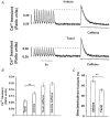
Figure 7. Effect of taxol on SR Ca2+ release
Ventricular myocytes were incubated in either 10 μM taxol or vehicle solution for 2 h at 37 °C and superfused with Tyrode solution. Initially cells were electrically stimulated at 1 Hz. Electrical stimulation was then turned off for 10 s and a switch to 10 mM caffeine was applied for 10 s. A, chart recordings showing the effects of caffeine on SR Ca2+ release in vehicle-treated (upper panel) and taxol-treated (lower panel) cells. B, amplitudes of Ca2+ transient (fura-2 ratio) during electrical stimulation or exposure to caffeine in taxol- or vehicle-treated cells. C, amplitude of Ca2+ transient during electrical stimulation expressed as a percentage of the Ca2+ transient during application of caffeine in vehicle- or taxol-treated cells. Values are means ±s.e.m.**P < 0·01 (n= 18).

References

    1. Arnal I, Wade RH. How does taxol stabilize microtubules? Current Biology. 1995;5:900–908. - PubMed
    1. Bassani JW, Yuan WL, Bers DM. Fractional SR Ca release is regulated by trigger Ca and SR Ca content in cardiac myocytes. American Journal of Physiology. 1995;37:C1313–1319. - PubMed
    1. Bers DM. Excitation-Contraction Coupling and Cardiac Contractile Force. Kluwer Academic Press; 1991.
    1. Brette F, Hongo K, Haroon MM, White E. Rapid negative inotropic effect of deuterium oxide in isolated ferret papillary muscles and single rat ventricular myocytes. The Journal of Physiology. 1996;491.P:157. P. - PubMed
    1. Cannell MB, Berlin JR, Lederer WJ. Effect of membrane potential changes on the calcium transient in single rat cardiac muscle cells. Science. 1987;238:1419–1423. - PubMed

Publication types

MeSH terms

LinkOut - more resources