. 1999 Apr 15;516 ( Pt 2)(Pt 2):525-38.
doi: 10.1111/j.1469-7793.1999.0525v.x.

Initiation of peristalsis by circumferential stretch of flat sheets of guinea-pig ileum

Affiliations

Initiation of peristalsis by circumferential stretch of flat sheets of guinea-pig ileum

S J Brookes et al. J Physiol. .

Abstract

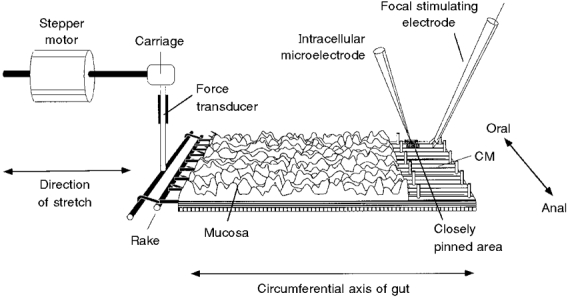
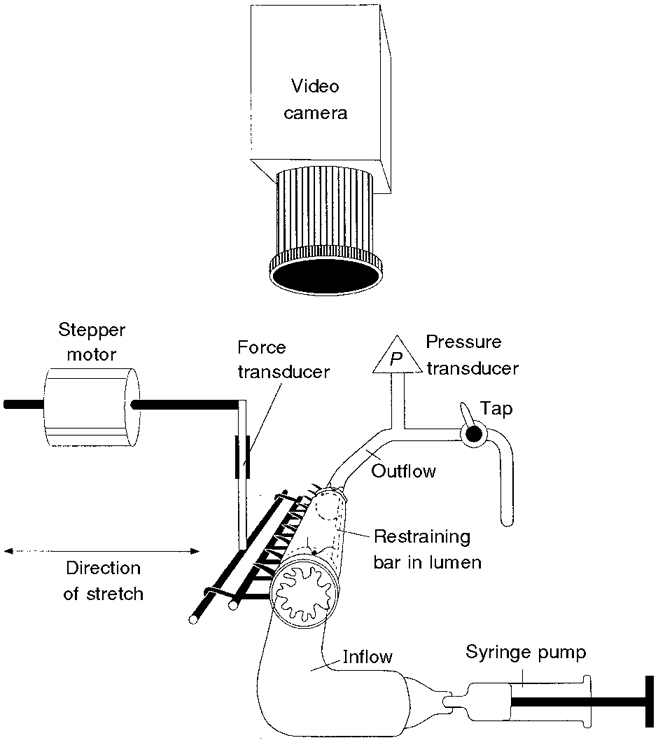
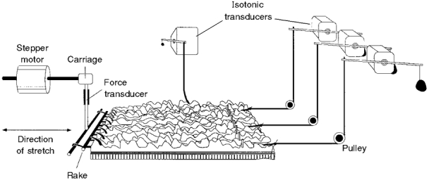
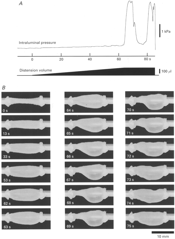
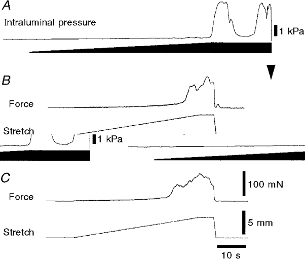
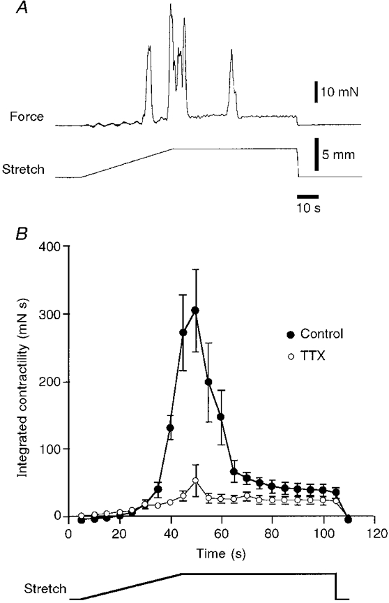
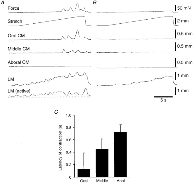
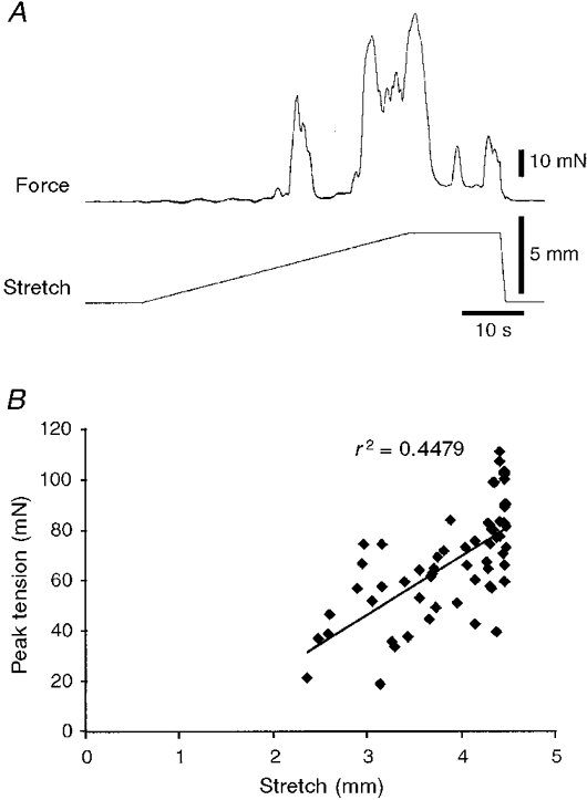
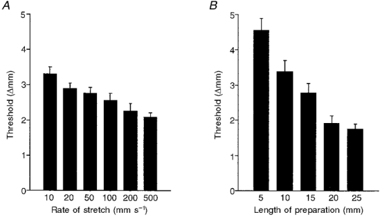
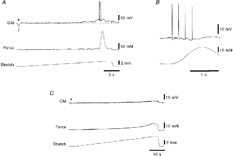
1. Segments of isolated guinea-pig intestine, 12 mm long, were distended slowly by intraluminal fluid infusion or by mechanical stretch as either a tube or flat sheet. In all cases, at a constant threshold length, a sudden, large amplitude contraction of the circular muscle occurred orally, corresponding to the initiation of peristalsis. 2. Circumferential stretch of flat sheet preparations evoked graded contractions of the longitudinal muscle (the 'preparatory phase'), which were maintained during circular muscle contraction. This suggests that the lengthening reported during the emptying phase of peristalsis is due to mechanical interactions. 3. The threshold for peristalsis was lower with more rapid stretches and was also lower in long preparations (25 mm) compared with short preparations (5-10 mm), indicating that ascending excitatory pathways play a significant role in triggering peristalsis. 4. Stretching a preparation beyond the threshold for peristalsis evoked contractions of increasing amplitude; thus peristalsis is graded above its threshold. However, during suprathreshold stretch maintained at a constant length, contractions of the circular muscle quickly declined in amplitude and frequency. 5. Circular muscle cells had a resting membrane potential approximately 6 mV more negative than the threshold for action potentials. During slow circumferential stretch, subthreshold graded excitatory motor input to the circular muscle occurred, prior to the initiation of peristalsis. However, peristalsis was initiated by a discrete large excitatory junction potential (12 +/- 2 mV) which evoked bursts of smooth muscle action potentials and which probably arose from synchronized firing of ascending excitatory neuronal pathways.


Figures

Figure 1
Figure 1. Flat sheet preparation used to study the effects of slow stretch on motor activity in the circular smooth muscle of the isolated guinea-pig ileum
One edge was pinned to the base of the organ bath and the other was connected via a claw, resembling a miniature garden rake, to an inflexible force transducer. The force transducer was mounted on the carriage of the tissue stretcher, which was driven by a stepper motor and used to stretch the preparation in the circumferential direction. In some preparations, the mucosa and submucosa were removed from the right-hand edge of the preparation, exposing the circular muscle (CM). A line of 50 μm diameter pins restricted movement of 2-3 mm of tissue along the pinned edge of the preparation and within this region 40-60 fine pins (made from 20 μm diameter tungsten wire) were placed in serried rows 140 μm apart to immobilize a small area of circular muscle. Care was taken to ensure that none of the pins was aligned circumferentially with the immobilized area, thus ensuring that this region remained functionally connected with the bulk of smooth muscle in the preparation. Conventional intracellular recordings were made from the immobilized area and a focal stimulating electrode was placed 1 mm circumferential to the pinned area to stimulate motor axons within the circular muscle.
Figure 2
Figure 2. Set-up used to stretch the same tubular preparation of guinea-pig ileum via infusion of Krebs solution into the lumen or by the tissue stretcher
A 12 mm length of ileum was ligated to the inflow and outflow cannulae (3 mm outer diameter), with the restraining bar (of approximately 1 mm outer diameter) running through the lumen. A miniature ‘rake’ was inserted though the wall of the intestine along its length and initially allowed to hang beneath the preparation. Krebs solution was infused into the lumen at 2·7 μl s−1 from a syringe pump until peristalsis was evoked. Intraluminal pressure (P) was monitored by a pressure transducer attached to the outflow cannula and the experiment was recorded with a video camera positioned above the preparation. After stable recordings had been obtained, the ligatures were cut and the ‘rake’ was hooked onto the transducer on the tissue stretcher. The preparation remained anchored by the restraining bar through the lumen and was stretched at 100 μm s−1 until a sudden contraction marked the initiation of peristalsis.
Figure 5
Figure 5. Preparation used to record circular and longitudinal muscle contractions at multiple points during slow circumferential stretch
The preparation was set up in a manner similar to that shown in Fig. 1 but without removal of the mucosa or submucosa. Four short notches (2 mm long) were cut into the immobilized edge of the preparation and the resulting peninsulas at the oral, middle and anal ends of the preparation, each 1-1·5 mm wide, were attached to isotonic transducers via a pulley, with a counterweight of 200 mg. The end of the preparation was also attached to an isotonic transducer by a long (30 cm) cotton thread to monitor longitudinal muscle contractions, with minimal interference from circular muscle movements.
Figure 3
Figure 3. Pressure and video recordings of a 12 mm preparation distended by infusion of fluid at 2·7 μl s−1
A shows the intraluminal pressure recording and a schematic representation of the infusion volume. In B, the appearance of the preparation at various times after the start of infusion (at 0 s) is shown on each image (oral is to the left). After the start of the infusion, intraluminal pressure increased gradually, corresponding to the slow distension of the preparation (from 0 to 60 s). Note that during this period there was a tendency for the aboral end of the preparation to be more distended than the oral end. At 64 s a substantial contraction of the circular muscle occurred at the oral end of the preparation, corresponding to the start of the pressure wave in A, and the syringe pump was switched off. The contraction increased in amplitude and spread along the preparation, displacing contents into the aboral region which became dramatically distended (expulsion of contents was prevented by the outflow tap being closed). By 70-72 s, the contraction started to wane and intraluminal pressure fell. A second wave of peristalsis started at 77-80 s. The outflow tap was opened at 86 s, draining the contents.
Figure 4
Figure 4. Peristalsis evoked in the same piece of tissue, as a tube and as a flat sheet
The effects of distension by slow fluid infusion (A), circumferential stretch of the intact tube with the tissue stretcher (B) and circumferential stretch of the flat sheet (C) are shown for the same segment of ileum. The trace in A is the same as that shown in Fig. 3A and the downward arrowhead marks the point at which the contents were drained from the preparation. Note that in each case, stretch evoked a small increase in wall tension (measured as pressure in A, and as force in B and C) until a threshold length, at which point there was a powerful contraction of the circular muscle, corresponding to the initiation of peristalsis. With fluid infusion maintained for 10 s (A), a second peristaltic pressure wave was recorded; similar findings were obtained with preparations stretched with the tissue stretcher, when suprathreshold lengths were maintained (see Fig. 7). Calibration bars in C also apply to B.
Figure 7
Figure 7. Effects of maintained, suprathreshold stretch
A, typical trace showing the effects of maintained suprathreshold stretch revealing a rapid run-down in peristaltic contractions. The threshold for peristalsis corresponded to a stretch of 3·0 mm. When the stretch was continued to 4·5 mm, another contraction of greater peak amplitude was evoked during the stretch. However, when the constant stretch of 4·5 mm was maintained, contraction amplitude declined, as did the frequency of contractions. B, mean contractile activity (measured as the integral of force over time, in 5 s time bins) plotted for 6 preparations (•), each stretched by 4·5 mm. It is clear that there was an increase in contractile activity after the threshold was reached, but that when a constant level of stretch was maintained, contractile activity declined rapidly. In the presence of 0·6 μM TTX (^) a lower level of muscle activity occurred throughout the maintained stretch, indicating that although peristaltic contractions disappeared during maintained stretch, there was still on-going excitatory neuronal input to the muscle.
Figure 6
Figure 6. Contractions of circular (CM) and longitudinal muscle (LM) evoked by slow circumferential stretch of a flat sheet preparation
A, the traces at the top show the slow ramp stretch (at 100 μm s−1 for approximately 3 mm) and the circular muscle force, showing the abrupt onset of contraction in the circular muscle. There was spontaneous contractile activity in the longitudinal muscle prior to stretch, which was enhanced during the stretch, and maintained during the initiation of peristalsis. The first area of circular muscle to contract was at the oral end of the preparation, with later and smaller contractions occurring further anally; in this particular run no contraction was recorded at the aboral end of the tissue. B, all contractile activity, in both circular and longitudinal muscle layers, was abolished by addition of 0·6 μM TTX to the bath for 6 min, although passive changes in length and tension of the muscle were still present, especially in the longitudinal muscle. The bottom trace in A shows the longitudinal muscle trace in B (in the presence of TTX) subtracted from the control response, reflecting the active, neuronally mediated response of the longitudinal muscle to stretch. C shows the mean latency of circular muscle contractions, relative to the initiation of peristalsis, recorded with the tissue stretcher for the oral, middle and anal ends of 4 preparations, showing that the oral site contracted, on average, before the other sites. In most cases the circular muscle contraction at the oral end of the preparation was largest and it diminished as it propagated along the preparation. In some cases, the aboral end of the tissue failed to contract at all - in these cases latency measurements could not be made and these traces were not included in the analysis.
Figure 8
Figure 8. Effects of stretching beyond the threshold for the initiation of peristalsis
A, slow circumferential stretch at 100 μm s−1 initiated peristalsis at a threshold length change of 2·6 mm. Continuing the stretch to 4·5 mm evoked two further major contractions of increasing amplitude. Typically, during the plateau phase of the stretch, subsequent contractions were reduced in amplitude and eventually disappeared altogether (see Fig. 7). B shows that there was a significant correlation between the peak tension of the phasic contractions evoked by stretch and the degree of stretch (n= 6). This indicates that although the first contraction occurs at a particular threshold length, peristalsis is not an all-or-nothing phenomenon, but rather is graded with increasing degrees of stretch.
Figure 9
Figure 9. Effects of rate of stretch and the length of the preparation on the threshold for peristalsis
Each graph shows combined data from 6 preparations; error bars represent s.e.m.A, less stretch was required to evoke peristalsis with faster rates of stretch indicating that there must be some run-down in the excitatory pathways prior to the initiation of peristalsis. B, shorter preparations (down to 5 mm in length) required more stretch to evoke circular muscle contractions than did longer preparations (up to 25 mm in length).
Figure 10
Figure 10. Intracellular recordings from circular muscle (CM) cells during slow, circumferential stretch
A, typical recording from a circular muscle cell located 2 mm from the oral end of a 12 mm long preparation showing the sudden contraction of the circular muscle after the preparation had been stretched 2·4 mm beyond its resting length of 12 mm. A single electrical stimulus (0·25 ms, 15 V) was applied via focal stimulating electrodes 1 mm circumferential to the recording site (asterisk) evoking a large inhibitory junction potential, confirming that the recording electrode was in a circular muscle cell. A small graded depolarization (about 3 mV in amplitude) occurred prior to the initiation of peristalsis (above the resting membrane potential shown by the dotted line). Just before the initiation of peristalsis there was a large compound excitatory junction potential (7 mV above resting membrane potential) with superimposed smooth muscle action potentials. In B, the same recording is shown on a faster time base, revealing that the burst of action potentials preceded the start of the contraction. This is consistent with the contractile activity appearing first at the oral end of the preparation close to where the recording was made. C, TTX (0·6 μM for 15 min) abolished the inhibitory junction potential (asterisk), and most electrical and contractile activity, leaving just a small myogenic depolarization of approximately 2 mV at the highest level of stretch. Note the different time scales in A, B and C and that the preparation was stretched 5 mm in C.

References

    1. Bayliss WM, Starling EH. The movements and innervation of the small intestine. The Journal of Physiology. 1899;24:99–143. - PMC - PubMed
    1. Bornstein JC, Costa M, Furness JB, Lang RJ. Electrophysiological analysis of projections of enteric inhibitory motoneurones in the guinea-pig small intestine. The Journal of Physiology. 1986;370:61–74. - PMC - PubMed
    1. Brookes SJH, Bywater RAR, Costa M. Characterisation of excitatory motor neurones to the circular muscle of the guinea-pig small intestine. Proceedings of the Australian Physiological and Pharmacological Society. 1995a;6:43.
    1. Brookes SJH, Meedeniya ACB, Jobling P, Costa M. Orally projecting interneurones in the guinea-pig small intestine. The Journal of Physiology. 1997;505:473–491. - PMC - PubMed
    1. Brookes SJH, Song ZM, Ramsay GA, Costa M. Long aboral projections of Dogiel type II, AH neurons within the myenteric plexus of the guinea pig small intestine. Journal of Neuroscience. 1995b;15:4013–4022. - PMC - PubMed

Publication types

LinkOut - more resources