Clinical Trial
. 1999 Apr 15;516 ( Pt 2)(Pt 2):549-57.
doi: 10.1111/j.1469-7793.1999.0549v.x.

Role of nitric oxide in the regulation of microvascular perfusion in human skin in vivo

Affiliations
Clinical Trial

Role of nitric oxide in the regulation of microvascular perfusion in human skin in vivo

G F Clough. J Physiol. .

Abstract

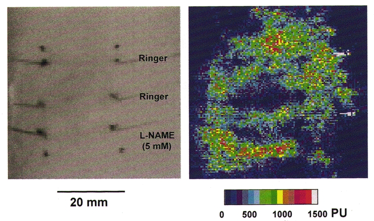
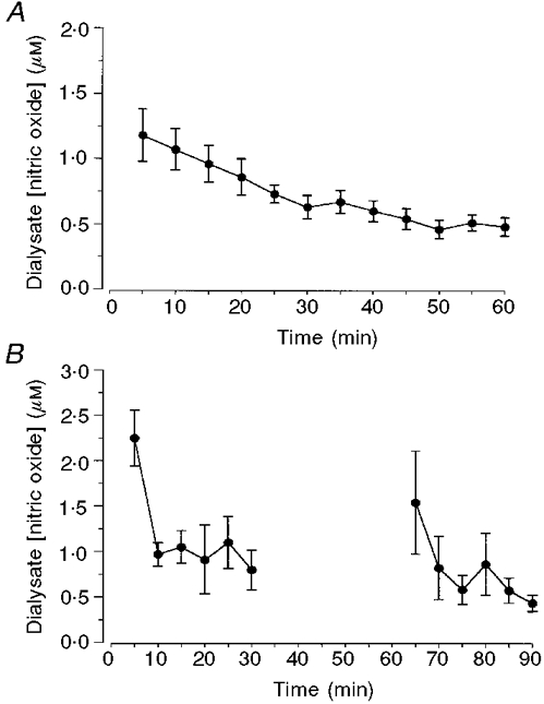
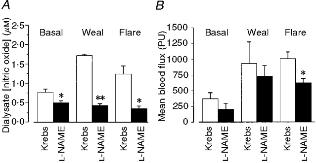
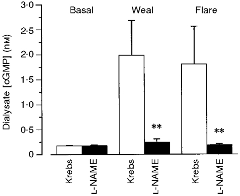
1. Nitric oxide (NO) concentrations were measured in dialysate from healthy human skin, in vivo, both at rest and during the inflammatory response to intradermal histamine or bradykinin. Changes in dialysate NO concentration, measured by electrochemical detection, were related to changes in dermal vascular perfusion, measured using scanning laser Doppler imaging. 2. Basal NO concentration in dermal microdialysate was 0.60 +/- 0.14 microM (mean +/- s.e.m.). Following the intradermal injection of histamine, a transient, time-dependent increase in NO concentration was measured in areas of skin incorporating the weal and in others incorporating the flare. The increase in NO concentration was associated with an increase in dialysate cGMP concentration in both the weal and flare areas. 3. Addition of N G-nitro-l-arginine-methyl ester (L-NAME, 5 mM) to the probe perfusate resulted in an inhibition of the histamine-induced increase in NO and cGMP. Moreover, the reduction in dialysate NO concentration was associated with a reduction in dermal vascular flux, both under basal conditions and within the weal and flare response. 4. These results demonstrate, by the use of microdialysis, that vasoactive mediators can be measured in healthy human skin in vivo. They provide direct evidence that endogenous concentration of NO increases during the inflammatory weal and flare response to histamine and that the increase in dermal NO concentration is associated with increases in cGMP concentration and dermal vascular perfusion, thus confirming a role for NO in vasoregulation in human skin.


Figures

Figure 1
Figure 1. Scanning laser Doppler image of the blood flux 10 min after intradermal injection of histamine
A high resolution scanning laser Doppler image of dermal blood flux taken 10 min after intradermal injection of histamine (15 μl of 1 μM). The injection was made adjacent to the central of three microdialysis probes inserted into the volar surface of the forearm. The upper two probes were perfused with Ringer solution and the lower one with L-NAME (5 mM). The same image is displayed as a grey scale image for the location of the fibres and as a coloured image used to calculate mean blood flux in a region 4 mm × 20 mm over each probe.
Figure 2
Figure 2. Effect of continued probe perfusion on dialysate NO concentration
A, dialysate concentration of NO was measured in 25 μl (5 min) samples collected during continuous perfusion of probes with Ringer solution (5 μl min−1). Data are means ±s.e.m. from 12 probes in 6 subjects. B, dialysate NO concentration was measured in 25 μl (6 × 5 min) samples collected during a 30 min period of continuous probe perfusion. Perfusion was then stopped and restarted 30 min later. Dialysate was collected for a further 30 min (6 × 5 min). Data are means ±s.e.m. of 4 probes in 1 subject.
Figure 3
Figure 3. NO release during the dermal vascular response to histamine or bradykinin
NO concentration in dialysate from Ringer solution-perfused probes inserted into the volar surface of the forearm of healthy volunteers. Agonists were injected close to 1 fibre, such that the weal developed over this fibre and the flare over a second inserted at a distance of 20 mm from the first. Dialysate samples (2 min, 10 μl) were collected before (•) and after (^) the intradermal injection of (A) histamine (15 μl of 1 μM) or (B) bradykinin (15 μl of 1 μM). All data represent the means ±s.e.m. from 9 subjects.
Figure 4
Figure 4. Peak NO release in response to intradermal histamine or vehicle control
Maximum values of dialysate NO concentration measured before (5) and after the injection of either histamine (15 μl of 1 μM) (▪) or vehicle control (formula image
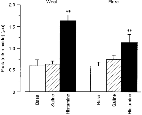
). Dialysate was collected from both the weal and flare areas of the histamine response. Maximum values in the weal occurred 2-4 min (P < 0·001) after provocation with histamine and that in the flare 6-8 min (P < 0·01). All columns represent the means +
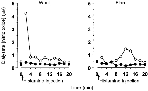
s.e.m. from 9 subjects.
Figure 5
Figure 5. Time course of the inhibition of histamine induced NO release by L-NAME
Data obtained from one subject from two pairs of dialysis probes inserted into the volar surface of one forearm (see Methods). Probes were perfused with either Ringer's solution (^) or Ringer's solution containing L-NAME (5 mM) (•) for 30 min prior to the injection of histamine (15 μl of 1 μM) close to one probe of each pair, such that the weal developed over one probe and the flare over the second. Dialysate was collected at 2 min intervals.
Figure 6
Figure 6. Effects of L-NAME on dialysate NO concentration and dermal vascular perfusion
A, L-NAME (5 mM) added to the microdialysis probe perfusate resulted in a fall in basal levels of NO (P < 0·03) (10 probes, 5 subjects). It reduced peak dialysate NO concentration following intradermal injection of histamine (15 μl of 1 μM) in both the weal (P < 0·007) and the flare response (P < 0·05). B, mean blood flux (in Moor LDI perfusion units, PU) measured in the region of the probe (see Fig. 1) was reduced by L-NAME in both resting skin (41 ± 10 %) and within the weal (17 ± 12 %) and flare (37 ± 5 %) response to histamine. The reduction only reached significance in the flare (P < 0·01). All columns represent means +s.e.m. from 5 subjects.
Figure 7
Figure 7. Effect of L-NAME on dialysate cGMP concentration following intradermal histamine
Dialysate cGMP concentration before and after the intradermal injection of histamine (15 μl of 1 μM) in the presence and absence of L-NAME (5 mM) added to the probe perfusate. The mean basal level of cGMP in the absence of L-NAME (0·18 ± 0·01 nM, 9 probes, 7 subjects) was not significantly different from that in its presence (0·17 ± 0·02 nM, 11 probes, 7 subjects). Addition of L-NAME to the perfusate significantly reduced the histamine-induced increase in cGMP concentration in both the weal and flare response (**P < 0·001) measured in adjacent sites on the forearms of 8 subjects.

Similar articles

Cited by

References

    1. Anderson C, Andersson T, Wårdell K. Changes in skin circulation after insertion of a microdialysis probe visualized by laser Doppler perfusion imaging. Journal of Investigative Dermatology. 1994;102:807–811. 10.1111/1523-1747.ep12378630. - DOI - PubMed
    1. Andoh T, Kuraishi Y. Quantitative determination of endogenous nitric oxide in the mouse skin in vivo by microdialysis. European Journal of Pharmacology. 1997;332:279–282. 10.1016/S0014-2999(97)01114-X. - DOI - PubMed
    1. Benrath J, Eschenfelder C, Zimmerman M, Gillardon F. Calcitonin gene-related peptide, substance P and nitric oxide are involved in cutaneous inflammation following ultraviolet irradiation. European Journal of Pharmacology. 1995;293:87–96. - PubMed
    1. Bito L, Dawson H, Levin E, Murray M, Snider N. The concentrations of free amino acids and other electrolytes in cerebral spinal fluid, in vivo dialysate of brain, and blood plasma of the dog. Journal of Neurological Chemistry. 1966;13:1057–1067. - PubMed
    1. Bruch-Gerharz D, Ruzicka T, Kolb-Bachofen V. Nitric oxide in human skin: current status and future prospects. Journal of Investigative Dermatology. 1998;110:1–7. - PubMed

Publication types

MeSH terms

LinkOut - more resources