

Expression of insulin growth factor-1 splice variants and structural genes in rabbit skeletal muscle induced by stretch and stimulation
- PMID: 10087355
- PMCID: PMC2269271
- DOI: 10.1111/j.1469-7793.1999.0583v.x
Expression of insulin growth factor-1 splice variants and structural genes in rabbit skeletal muscle induced by stretch and stimulation
Abstract
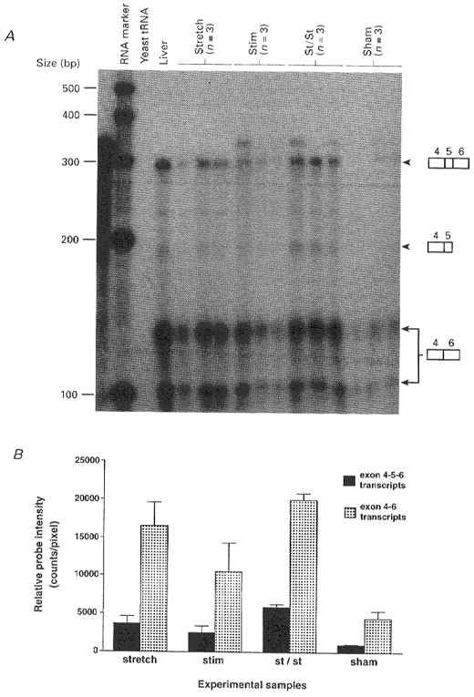
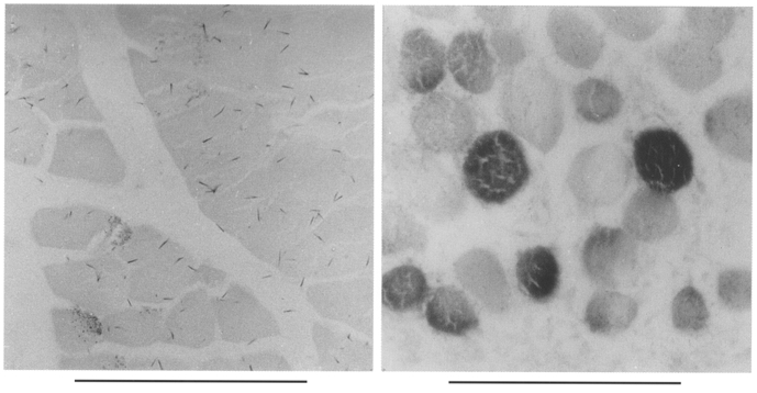
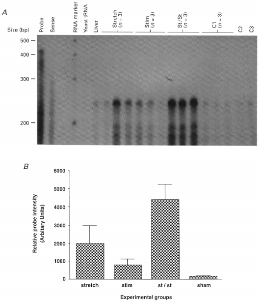
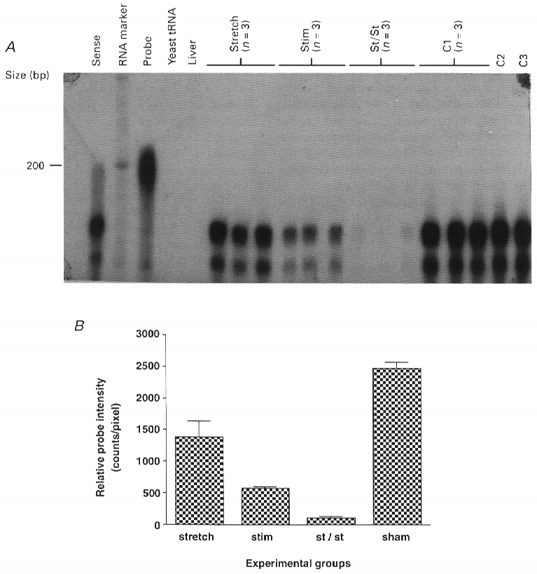
1. Skeletal muscle is a major source of circulating insulin growth factor-1 (IGF-1), particularly during exercise. It expresses two main isoforms. One of the muscle IGF-1 isoforms (muscle L.IGF-1) is similar to the main liver IGF-1 and presumably has an endocrine action. The other muscle isoform as a result of alternative splicing has a different 3' exon sequence and is apparently designed for an autocrine/paracrine action (mechano-growth factor, MGF). Using RNase protection assays with a probe that distinguishes these differently spliced forms of IGF-1, their expression and also the expression of two structural genes was measured in rabbit extensor digitorum longus muscles subjected to different mechanical signals. 2. Within 4 days, stretch using plaster cast immobilization with the limb in the plantar flexed position resulted in marked upregulation of both forms of IGF-1 mRNA. Electrical stimulation at 10 Hz combined with stretch (overload) resulted in an even greater increase of both types of IGF-1 transcript, whereas electrical stimulation alone, i.e. without stretch, resulted in no significant increase over muscle from sham-operated controls. Previously, it was shown that stretch combined with electrical stimulation of the dorsiflexor muscles in the adult rabbit results in a marked increase in muscle mass involving increases in both length and girth, within a few days. The expression of both systemic and autocrine IGF-1 growth factors provides a link between the mechanical signal and the marked increase in the structural gene expression involved in tissue remodelling and repair. 3. The expression of the beta actin gene was seen to be markedly upregulated in the stretched and stretched/stimulated muscles. It was concluded that the increased expression of this cytoskeletal protein gene is an indication that the production of IGF-1 may initially be a response to local damage. 4. Switches in muscle fibre phenotype were studied using a specific gene probe for the 2X myosin heavy chain gene. Type 2X expression was found to decrease markedly with stimulation alone and when electrical stimulation was combined with stretch. Unlike the induction of IGF-1 and beta actin, the decreased expression of the 2X myosin mRNA was less marked in the 'stretch only' muscles. This indicates that the interconversion of fibre type 2X to 2A may in some situations be commensurate with, but not under the control of IGF-1.
Figures
Similar articles
-
Changes in muscle mass and phenotype and the expression of autocrine and systemic growth factors by muscle in response to stretch and overload.J Anat. 1999 Apr;194 ( Pt 3)(Pt 3):323-34. doi: 10.1046/j.1469-7580.1999.19430323.x. J Anat. 1999. PMID: 10386770 Free PMC article. Review.
-
Changes in muscle fibre type, muscle mass and IGF-I gene expression in rabbit skeletal muscle subjected to stretch.J Anat. 1997 May;190 ( Pt 4)(Pt 4):613-22. doi: 10.1046/j.1469-7580.1997.19040613.x. J Anat. 1997. PMID: 9183683 Free PMC article.
-
Expression and splicing of the insulin-like growth factor gene in rodent muscle is associated with muscle satellite (stem) cell activation following local tissue damage.J Physiol. 2003 Jun 1;549(Pt 2):409-18. doi: 10.1113/jphysiol.2002.035832. Epub 2003 Apr 11. J Physiol. 2003. PMID: 12692175 Free PMC article.
-
Gene expression in muscle in response to exercise.J Muscle Res Cell Motil. 2003;24(2-3):121-6. doi: 10.1023/a:1026041228041. J Muscle Res Cell Motil. 2003. PMID: 14609023 Review.
-
Cloning and characterization of an IGF-1 isoform expressed in skeletal muscle subjected to stretch.J Muscle Res Cell Motil. 1996 Aug;17(4):487-95. doi: 10.1007/BF00123364. J Muscle Res Cell Motil. 1996. PMID: 8884603
Cited by
-
Stimulation of mechano-growth factor expression by myofibrillar proteins in murine myoblasts and myotubes.Mol Cell Biochem. 2012 Apr;363(1-2):347-55. doi: 10.1007/s11010-011-1187-5. Epub 2011 Dec 9. Mol Cell Biochem. 2012. PMID: 22160926
-
Muscle growth: no IGFs, ands, or buts.J Physiol. 2008 Jan 1;586(1):5-6. doi: 10.1113/jphysiol.2007.147660. J Physiol. 2008. PMID: 18167368 Free PMC article. No abstract available.
-
Functional deficits and insulin-like growth factor-I gene expression following tourniquet-induced injury of skeletal muscle in young and old rats.J Appl Physiol (1985). 2008 Oct;105(4):1274-81. doi: 10.1152/japplphysiol.90418.2008. Epub 2008 Jul 31. J Appl Physiol (1985). 2008. PMID: 18669936 Free PMC article.
-
The Role of Nutri(epi)genomics in Achieving the Body's Full Potential in Physical Activity.Antioxidants (Basel). 2020 Jun 7;9(6):498. doi: 10.3390/antiox9060498. Antioxidants (Basel). 2020. PMID: 32517297 Free PMC article. Review.
-
Insulin-like growth factor I (IGF-1) Ec/Mechano Growth factor--a splice variant of IGF-1 within the growth plate.PLoS One. 2013 Oct 11;8(10):e76133. doi: 10.1371/journal.pone.0076133. eCollection 2013. PLoS One. 2013. PMID: 24146828 Free PMC article.
References
-
- Barton-Davis ER, LaFramoise WA, Kushmerick MJ. Activity-dependent induction of slow myosin gene expression in isolated fast-twitch mouse muscle. American Journal of Physiology. 1996;271:C1409–1414. - PubMed
-
- Booth FW, Thomason DB. Molecular and cellular adaptation of muscle in response to exercise: perspectives of various models. Physiological Reviews. 1991;71:541–585. - PubMed
-
- Brahm H, Piehl-Aulin K, Saltin B, Ljunghall S. Net fluxes over working thigh of hormones, growth factors and biomarkers of bone metabolism during lasting dynamic exercise. Calcified Tissue. 1997;60:175–180. - PubMed
-
- Chew SL, Lavender P, Clark AJL, Ross RJM. An alternative spliced human insulin-like growth transcript with hepatic tissue expression that diverts away from the mitogenic IBPE1 peptide. Endocrinology. 1995;136:1939–1944. - PubMed
Publication types
MeSH terms
Substances
Grants and funding
LinkOut - more resources
Full Text Sources
Other Literature Sources
Miscellaneous