. 1999 Apr 2;274(14):9607-16.
doi: 10.1074/jbc.274.14.9607.

Human pancreatic islets express mRNA species encoding two distinct catalytically active isoforms of group VI phospholipase A2 (iPLA2) that arise from an exon-skipping mechanism of alternative splicing of the transcript from the iPLA2 gene on chromosome 22q13.1

Affiliations

Human pancreatic islets express mRNA species encoding two distinct catalytically active isoforms of group VI phospholipase A2 (iPLA2) that arise from an exon-skipping mechanism of alternative splicing of the transcript from the iPLA2 gene on chromosome 22q13.1

Z Ma et al. J Biol Chem. .

Abstract

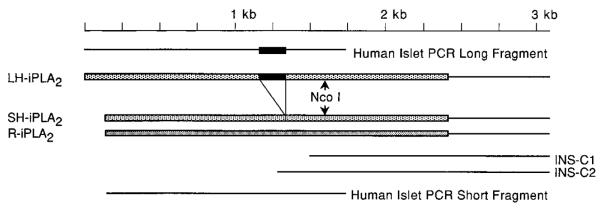
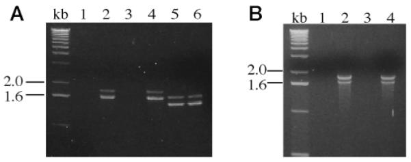
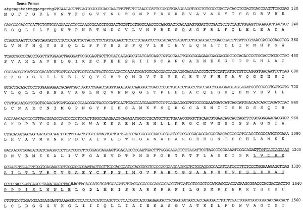
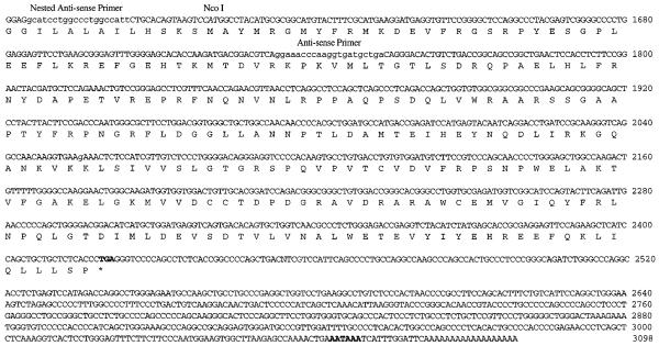
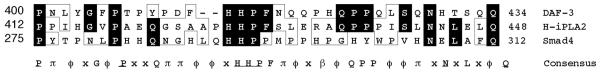
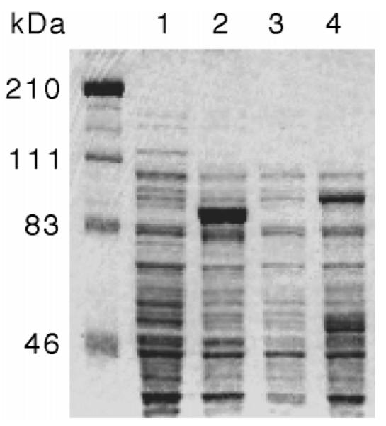
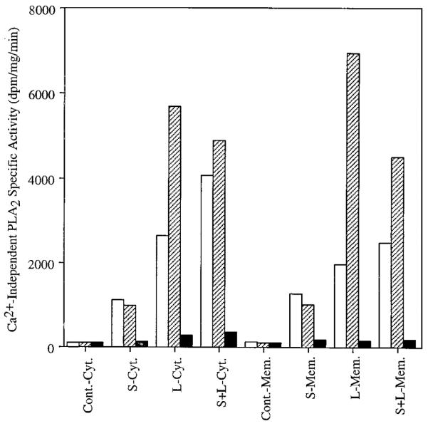
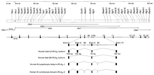
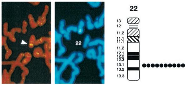
An 85-kDa Group VI phospholipase A2 enzyme (iPLA2) that does not require Ca2+ for catalysis has recently been cloned from three rodent species. A homologous 88-kDa enzyme has been cloned from human B-lymphocyte lines that contains a 54-amino acid insert not present in the rodent enzymes, but human cells have not previously been observed to express catalytically active iPLA2 isoforms other than the 88-kDa protein. We have cloned cDNA species that encode two distinct iPLA2 isoforms from human pancreatic islet RNA and a human insulinoma cDNA library. One isoform is an 85-kDa protein (short isoform of human iPLA2 (SH-iPLA2)) and the other an 88-kDa protein (long isoform of human iPLA2 (LH-iPLA2)). Transcripts encoding both isoforms are also observed in human promonocytic U937 cells. Recombinant SH-iPLA2 and LH-iPLA2 are both catalytically active in the absence of Ca2+ and inhibited by a bromoenol lactone suicide substrate, but LH-iPLA2 is activated by ATP, whereas SH-iPLA2 is not. The human iPLA2 gene has been found to reside on chromosome 22 in region q13.1 and to contain 16 exons represented in the LH-iPLA2 transcript. Exon 8 is not represented in the SH-iPLA2 transcript, indicating that it arises by an exon-skipping mechanism of alternative splicing. The amino acid sequence encoded by exon 8 of the human iPLA2 gene is proline-rich and shares a consensus motif of PX5PX8HHPX12NX4Q with the proline-rich middle linker domains of the Smad proteins DAF-3 and Smad4. Expression of mRNA species encoding two active iPLA2 isoforms with distinguishable catalytic properties in two different types of human cells demonstrated here may have regulatory or functional implications about the roles of products of the iPLA2 gene in cell biologic processes.


Figures

Fig. 1
Fig. 1. Summary of cDNA fragments used to construct cDNA species containing the complete coding sequences of human islet iPLA2 isoforms
The two cDNA clones obtained by screening the human insulinoma cell cDNA library that contain the 3′-sequence of the human islet iPLA2 cDNA are designated INS-C1 and INS-C2. The RT-PCR products obtained using human islet RNA as template that contain the 5′-end of the human islet iPLA2 coding sequence (see Fig. 2) are designated human islet PCR long fragment and human islet PCR short fragment. Arrows indicate the location of the recognition site for the restriction endonuclease NcoI that is contained in the region of overlap between the insulinoma cell cDNA fragments and the human islet RNA RT-PCR products. The cDNA species containing the complete coding sequence of the long and short isoforms of human iPLA2 are designated LH-iPLA2 and SH-iPLA2, respectively. These cDNA species were prepared by NcoI digestion and ligation of the insulinoma cell cDNA fragment and one of the two RT-PCR products derived from human islet RNA. R-iPLA2, rat islet iPLA2 cDNA. The region of the 162-bp insert that distinguishes LH-iPLA2 from SH-iPLA2 is indicated by the black bar. The remainder of the coding sequence is indicated by shaded bars. The lighter shading represents human iPLA2 coding sequence; the darker shading represents rat iPLA2 coding sequence.
Fig. 2
Fig. 2. Agarose gel electrophoretic analyses of products of RT-PCRs performed with human islet RNA or U937 cell RNA as template and primers designed to amplify the 5′-end of human iPLA2 cDNA
RT-PCRs were performed with RNA isolated from human islets from two different donors (A) or from two different preparations of human promonocytic U937 cell RNA (B). In A, experiments shown in lanes 1, 2, and 5 were performed with RNA from islets from donor 1, and experiments shown in lanes 3, 4, and 6 were performed with RNA from islets from donor 2. Reverse transcriptase was omitted from the reactions analyzed in lanes 1 and 3 to exclude contamination from genomic DNA in the human islet RNA preparations. In reactions analyzed in lanes 1–4 (A), a set of PCR primers was used that was expected to yield a single 1.65-kb product, based on the rat islet iPLA2 cDNA sequence. In reactions analyzed in lanes 5 and 6 (A), the same 5′-primer was used as in the reactions analyzed in lanes 1–4, but a different 3′-primer was used that was expected to yield a shorter product, based on the rat iPLA2 cDNA sequence. The sequences of the 5′-primer and of the two 3′-primers used in these reactions are specified under “Experimental Procedures.” In B, experiments shown in lanes 1 and 2 were performed with RNA from U936 cell preparation 1, and experiments shown in lanes 3 and 4 were performed with RNA from U937 cell preparation 2. Reverse transcriptase was omitted from the reactions analyzed in lanes 1 and 3 (B). In reactions analyzed in lanes 1–4 (B), the set of PCR primers was the same as that in lanes 1–4 of A. Both of the RT-PCR products visualized in lanes 2 and 4 (B) were subcloned and sequenced, and the results were identical to those for the products in lanes 2 and 4 of A.
Fig. 3
Fig. 3. Nucleotide and deduced amino acid sequences of cDNA species encoding the long and short isoforms of human iPLA2
Species of cDNA containing the full coding region and the 3′-untranslated region for transcripts encoding LH-iPLA2 and SH-iPLA2 were constructed with cDNA fragments from the INS-C1 and INS-C2 clones and from the long and short fragments obtained from RT-PCRs with human islet RNA, as illustrated schematically in Fig. 1. The resultant cDNA species were subcloned and sequenced. The figure displays the nucleotide (top row) and deduced amino acid sequences (bottom row) for the LH-iPLA2 cDNA. The sequences of the forward primer, the reverse primer, and the nested reverse primer used in the RT-PCRs with human islet RNA are indicated, as is the location of the NcoI restriction endonuclease site. The 162-bp insert that is present in LH-iPLA2 cDNA but absent from SH-iPLA2 cDNA is underlined. The stop codon TGA is indicated by an asterisk. The presumptive polyadenylation signal sequence is indicated in boldface type.
Fig. 4
Fig. 4. Comparison of the amino acid sequence of the proline-rich insert in the long form of human iPLA2 to sequences in the proline-rich middle linker domains of the Smad proteins DAF-3 and Smad4
The deduced amino acid sequence between residues 412–448 for the long isoform of human iPLA2 is designated H-iPLA2 in the figure. A BLAST search indicated similarities between this sequence and that of residues 400–434 of the C. elegans DAF-3 Smad protein, which falls within the proline-rich middle linker domain of that protein. DAF-3 is most closely related to the mammalian protein Smad4, and residues 275–312 within the proline-rich middle linker domain of the Smad4 sequence are illustrated in the figure. Amino acid residues that are contained in the iPLA2 sequence and one or both of the other sequences are illustrated by dark boxes, and residues with chemically similar side chains are illustrated by light boxes. In the consensus sequence, residues that are identical among the three sequences are indicated by underlined, capitalized Roman letters. Residues that are common to the iPLA2 sequence and to one but not both of the other sequences are indicated by capitalized Roman letters that are not underlined. Positions at which residues with chemically similar side chains are observed in two or three of the sequences are designated with Greek letters. Acidic residues (Asp and Glu) are denoted by α; basic residues (His, Lys, and Arg) by b; neutral, nonpolar residues (Ala, Phe, Ile, Leu, Met, Pro, Val, and His) by ϕ; and neutral, polar residues (Gly, Asn, Gln, Ser, Thr, and Tyr) by p. Positions at which there is no similarity among the three sequences are denoted with an x.
Fig. 5
Fig. 5. Bacterial expression of the long isoform and short isoform human islet iPLA2 proteins
The cDNA species containing the complete coding regions of the short isoform (lanes 1 and 2) or the long isoform (lanes 3 and 4) of human islet iPLA2 were subcloned into the bacterial expression vector pET-28c (Novagen). The pET28-iPLA2 constructs were then used to transform the bacterial expression host BL21(DE3) (Novagen). Expression of proteins encoded by the cDNA inserts was induced by treating the cells with IPTG, and proteins expressed by induced (lanes 2 and 4) and noninduced (lanes 1 and 3) cells were compared by SDS-polyacrylamide gel electrophoresis analyses with Coomassie Blue staining. The expected molecular mass of the short isoform of human islet iPLA2 is 85 kDa (lane 2), and that of the long isoform of human islet iPLA2 is 88 kDa (lane 4).
Fig. 6
Fig. 6. Catalytic activities of recombinant long and short isoforms of human islet iPLA2 expressed in Sf9 cells
Recombinant baculovirus that contained cDNA inserts encoding either the long or short isoforms of human islet iPLA2 were prepared and used to infect Sf9 cells. At 48 h after infection, subcellular fractions were prepared from the Sf9 cells and assayed for iPLA2 activity with a radiolabeled phospholipid substrate. Uninfected Sf9 cells exhibited no detectable iPLA2 activity in either cytosol (Cont.-Cyt.) or membranes (Cont.-Mem.). Activity was observed in both cytosolic (S-Cyt. and L-Cyt.) and membranous (S-Mem. and L-Mem.) fractions of cells infected with baculovirus that contained cDNA inserts encoding LH-iPLA2 or SH-iPLA2, and activity was also observed in both subcellular fractions when cells were co-infected with baculovirus mixtures that contained both the LH-iPLA2 and SH-iPLA2 cDNA inserts (S+L-Cyt. and S+L-Mem.). The iPLA2 activities expressed in cells infected with baculovirus that contained cDNA encoding either human islet iPLA2 isoform were susceptible to inhibition by the iPLA2 suicide substrate BEL (filled bars). ATP (1 mM) was included in some assay solutions (hatched bars). Open bars, no ATP.
Fig. 7
Fig. 7. Schematic representation of the structure of the human iPLA2 gene and its restriction endonuclease map
The line at the top of the diagram indicates the scale in kb. There is an interruption in the scale between 0 and 25 kb because of the long length of the first intron. The locations of cleavage sites for restriction endonucleases are illustrated beneath the scale line. Below the summary of endonuclease sites, the lines designated HG6, HG5, HG4, HG7, HG3, and HG8 represent the regions of sequence contained in separate genomic clones (HGn) obtained from screening a human genomic library with 32P-labeled cDNA containing the full coding sequence of the long isoform of human islet iPLA2 and the 3′-untranslated region of its transcript. The genomic clones span over 60 kb of DNA and contain 16 exons represented in the cDNA for the long isoform of human iPLA2 that include 5′-untranslated region, the entire coding region, and 3′-untranslated region. The line below the genomic clone lines represents the locations of exons, which are identified by black rectangles, and the approximate lengths of the intervening introns are indicated by the lengths of the lines between the exons. The dark portions of the rectangles representing the exons indicate coding regions, and the unshaded portions of the rectangles representing exons 1 and 16 indicate untranslated regions. In the lower portion of the diagram, the region of the gene that includes exons 7–10 and the intervening introns is represented on an expanded scale, and the number of base pairs in each exon in this region is indicated. The regions of the gene that are included in four recognized splice-variant products iPLA2 gene are illustrated schematically in the bottom four lines. The human islet LH-iPLA2 isoform transcript contains exons 1–16 but not the alternate exons 8b and 9b. The human islet SH-iPLA2 isoform transcript contains exons 1–7 and 9–16 but not exon 8 or alternate exons 8b or 9b. Two iPLA2 splice variants have been reported by others in human B-lymphocyte lines (21). The transcripts for these variants contain intron sequences that result in premature stop codons and encode truncated forms of iPLA2 that contain the ankyrin repeat domain but lack the catalytic domain. These variants are designated human B-lymphocyte ankyrin-iPLA2-1 and human B-lymphocyte ankyrin-iPLA2-2. The open rectangles reflect the sites of the intron sequences that are contained in these truncation variants, and the location of these intron sequences in the iPLA2 gene are designated by sites 8b and 9b.
Fig. 8
Fig. 8. Localization of the human iPLA2 gene to chromosome 22q13.1 by fluorescence in situ hybridization
A genomic clone identified in screening the human genomic DNA library with the iPLA2 cDNA was biotinylated to generate a probe for FISH experiments with human lymphocyte chromosomes. Using this probe, 91 of 100 examined mitotic figures exhibited fluorescent signals on one pair of chromosomes. The white arrowhead in the left panel indicates the location of the two intense fluorescent spots reflecting hybridization of the probe with each member of the chromosome pair. The center panel illustrates the DAPI staining pattern of the same mitotic figure and identifies the chromosome as number 22. The diagram in the right panel illustrates regions of human chromosome 22 determined by DAPI banding patterns. These patterns were correlated with the site of fluorescent signal from the biotinylated iPLA2 genomic clone. A detailed positional assignment was achieved from analyses of 10 photographs from different preparations. The black circles indicate the location of the probe observed in each of the 10 experiments. In each case, the probe localized to region q13.1 of human chromosome 22. No other loci of hybridization of the biotinylated probe were observed.

References

    1. Dennis EA. J. Biol. Chem. 1994;269:13057–13060. - PubMed
    1. Gijon MA, Leslie CC. Cell Dev. Biol. 1997;8:297–303. - PubMed
    1. Dennis EA. Trends Biochem. Sci. 1997;22:1–2. - PubMed
    1. Balsinde J, Dennis EA. J. Biol. Chem. 1997;272:16069–16072. - PubMed
    1. Leslie CC. J. Biol. Chem. 1997;272:16709–16712. - PubMed

Publication types

MeSH terms