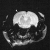
Comparison of ultrasmall particles of iron oxide (USPIO)-enhanced T2-weighted, conventional T2-weighted, and gadolinium-enhanced T1-weighted MR images in rats with experimental autoimmune encephalomyelitis
- PMID: 10094342
- PMCID: PMC7056125
Comparison of ultrasmall particles of iron oxide (USPIO)-enhanced T2-weighted, conventional T2-weighted, and gadolinium-enhanced T1-weighted MR images in rats with experimental autoimmune encephalomyelitis
Abstract
Background and purpose: Ultrasmall particles of iron oxide (USPIO) constitute a contrast agent that accumulates in cells from the mononuclear phagocytic system. In the CNS they may accumulate in phagocytic cells such as macrophages. The goal of this study was to compare USPIO-enhanced MR images with conventional T2-weighted images and gadolinium-enhanced T1-weighted images in a model of experimental autoimmune encephalomyelitis (EAE).
Methods: Nine rats with EAE and four control rats were imaged at 4.7 T and 1.5 T with conventional T1- and T2-weighted sequences, gadolinium-enhanced T1-weighted sequences, and T2-weighted sequences obtained 24 hours after intravenous injection of a USPIO contrast agent, AMI-227. Histologic examination was performed with hematoxylin-eosin stain, Perls' stain for iron, and ED1 immunohistochemistry for macrophages.
Results: USPIO-enhanced images showed a high sensitivity (8/9) for detecting EAE lesions, whereas poor sensitivity was obtained with T2-weighted images (1/9) and gadolinium-enhanced T1-weighted images (0/9). All the MR findings in the control rats were negative. Histologic examination revealed the presence of macrophages at the site where abnormalities were seen on USPIO-enhanced images.
Conclusion: The high sensitivity of USPIO for macrophage activity relative to other imaging techniques is explained by the histologic findings of numerous perivascular cell infiltrates, including macrophages, in EAE. This work supports the possibility of intracellular USPIO transport to the CNS by monocytes/macrophages, which may have future implications for imaging of human inflammatory diseases.
Figures


Comment in
-
Imaging macrophage activity in the brain by using ultrasmall particles of iron oxide.AJNR Am J Neuroradiol. 2000 Oct;21(9):1767-8. AJNR Am J Neuroradiol. 2000. PMID: 11039364 No abstract available.
References
-
- Weissleder R, Cheng H-C, Bogdanova A, Bogdanov A Jr. Magnetically labeled cells can be detected by MR imaging. J Magn Reson Imaging 1997;7:258-263 - PubMed
-
- Pouliquen D, Le Jeune JJ, Perdrisot R, Ermias A, Jallet P. Iron oxide nanoparticles for use as MRI contrast agent: pharmacokinetics and metabolism. Magn Reson Imaging 1991;9:275-283 - PubMed
-
- Anzai Y, Blackwell KE, Hirschowitz SL, et al. Initial clinical experience with dextran-coated superparamagnetic iron oxide for detection of lymph node metastases in patients with head and neck cancer. Radiology 1994;192:709-715 - PubMed
-
- Zimmer C, Weissleder R, Poss K, Bogdanova A, Carter Wright S, Scott Enochs W. MR imaging of phagocytosis in experimental gliomas. Radiology 1995;197:533-538 - PubMed
-
- Dousset V, Ballarino L, Delalande C, et al. Cerebral macrophage activity imaging using USPIO (abstr). In: Proceedings of the 35th Annual Meeting of the American Society of Neuroradiology, Toronto, 1997 Oak Brook, IL: American Society of Neuroradiology; 1997:70
Publication types
MeSH terms
Substances
LinkOut - more resources
Full Text Sources
Other Literature Sources
Medical
