

Use of three-dimensional MR angiography for tracking a contrast bolus in the carotid artery
Affiliations
- PMID: 10094349
- PMCID: PMC7056113
Item in Clipboard
Use of three-dimensional MR angiography for tracking a contrast bolus in the carotid artery
AJNR Am J Neuroradiol.
1999 Feb.
Abstract
Contrast-bolus tracking in the carotid bifurcation was accomplished using an MR angiographic technique with a 3D turbo field-echo readout (TR/TE = 6/3, flip angle = 50 degrees) modified by a keyhole scheme. Optimal visibility of the contrast bolus was achieved by digital subtraction from a reference volume. This technique reliably time-resolves the carotid arteries from the jugular veins.
Figures
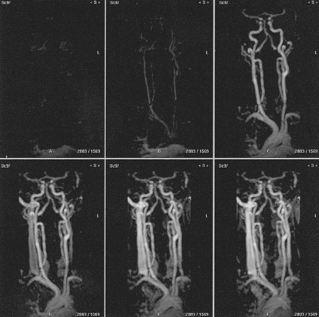
MIP projections of six of 20 temporally resolved volumes acquired from a male volunteer using subtracted 3D dynamic keyhole turbo field-echo MR angiography (TR/TE = 6/3, flip angle = 50°) show a coronal view of the vessels in the head and neck. The early phases show the aortic arch and major branches, the carotid arteries and bifurcations, and the vertebrobasilar arteries without interference from veins. The later phases show the arteries and the internal jugular veins
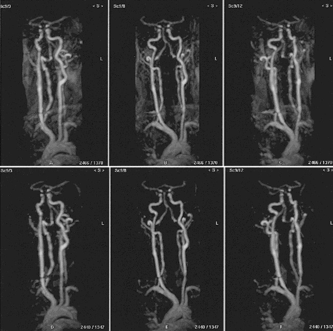
Rotated MIP projections of the eighth dynamic volume (arterial phase) acquired from a male volunteer using 3D dynamic keyhole turbo field-echo MR angiography (TR/TE = 6/3, flip angle = 50°). The nonsubtracted (top row) and the subtracted (bottom row) images show the aortic arch and major branches, the carotid arteries and bifurcations, and the vertebrobasilar arteries without interference from veins. Note the improved contrast between the arteries and background on the subtracted images
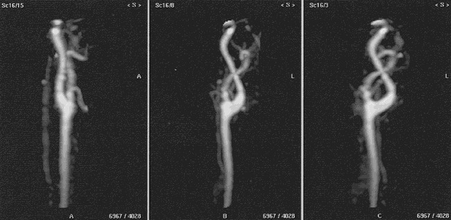
Rotated targeted MIP projections of the ninth dynamic volume (arterial phase) acquired from a female volunteer using subtracted 3D dynamic keyhole turbo field-echo MR angiography (TR/TE = 6/3, flip angle = 50°) show a normal left carotid bifurcation without interference from veins.
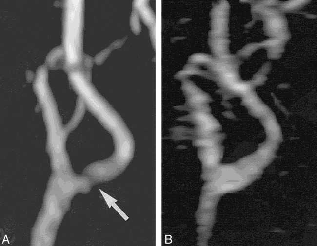
A, MIP projection of the left carotid bifurcation acquired from a female patient with suspected moderate narrowing (arrow) using 2D TOF MR angiography (TR/TE = 22/7, flip angle = 60°). B, The MIP projection acquired using subtracted 3D dynamic keyhole turbo field-echo MR angiography (TR/TE = 6/3, flip angle = 50°) shows a patent left carotid bifurcation.
Comment in
-
Assessing effective transvascular particle delivery to the brain parenchyma: a challenge to neuroradiology.AJNR Am J Neuroradiol. 1999 Feb;20(2):186. AJNR Am J Neuroradiol. 1999. PMID: 10094333 Free PMC article. No abstract available.
References
-
- Mayberg MR, Winn HR. Endarterectomy for asymptomatic carotid artery stenosis: resolving the controversy. JAMA 1995;273:1459-1461 - PubMed
-
- Masaryk AM, Ross JS, DiCello MC, et al. 3DFT MR angiography of the carotid bifurcation: potential and limitations as a screening examination. Radiology 1991;179:797-804 - PubMed
-
- Blatter DD, Bahr AL, Parker DL, et al. Cervical carotid MR angiography with multiple overlapping thin-slab acquisition: comparison with conventional angiography. AJR Am J Roentgenol 1993;161:1269-1277 - PubMed
-
- Urchuk SN, Plewes DB. Mechanisms of flow-induced signal loss in MR angiography. J Magn Reson Imaging 1992;2:453-462 - PubMed
-
- Lin W, Haake EM, Smith AS. Lumen definition in MR angiography.. J Magn Reson Imaging 1991;1:327-336 - PubMed
Publication types
MeSH terms
Substances
LinkOut - more resources
Full Text Sources