Review
. 1999 Feb;20(2):316-23.

Globoid cell leukodystrophy: distinguishing early-onset from late-onset disease using a brain MR imaging scoring method

Affiliations
Review

Globoid cell leukodystrophy: distinguishing early-onset from late-onset disease using a brain MR imaging scoring method

D J Loes et al. AJNR Am J Neuroradiol. 1999 Feb.

Abstract

Background and purpose: Our purpose was to determine the characteristic MR features of early-onset (before age 2 years) versus late-onset (after age 2 years) globoid cell leukodystrophy (GLD).

Methods: Thirty-four brain MR images in 22 patients with GLD were reviewed. A severity score (0 to 32), based on a point system derived from the location and extent of disease and the presence of focal and/or global atrophy, was calculated for each examination.

Results: Of the 22 patients, three were asymptomatic and 19 were symptomatic. Ten patients had early-onset disease, whereas nine had late-onset disease. MR images of all patients showed abnormalities. In the early-onset group (n = 10; mean maximum MR score, 8.1; range, 3-18), 90% had pyramidal tract involvement, 80% had cerebellar white matter involvement, 70% had deep gray matter involvement, 60% had posterior corpus callosal involvement, 50% had parietooccipital white matter involvement, and 40% had cerebral atrophy. Serial MR imaging in four of these patients revealed progressive disease. In the late-onset group (n = 9; mean maximum MR score, 5.6; range, 4-10), 100% had pyramidal tract involvement, 100% had parietooccipital white matter involvement, 89% had posterior corpus callosal involvement, and none had cerebellar white matter involvement, deep gray matter involvement, or cerebral atrophy. Serial MR imaging in one patient with late-onset GLD did not reveal any change. A spectrum of findings was observed in the three patients who were asymptomatic.

Conclusion: Cerebellar white matter and deep gray matter involvement are present only in early-onset GLD. Pyramidal tract involvement is a characteristic finding in both early- and late-onset GLD. This scoring method for brain MR observations will assist in the objective assessment of the impact of hematopoietic stem cell transplantation in patients with GLD.


Figures

<sc>fig</sc> 1.
fig 1.
GLD MR severity score versus age. Connecting lines indicate patients with serial examinations
<sc>fig</sc> 2.
fig 2.
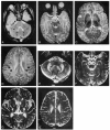
Patient 1: early-onset GLD. A–D, Axial T2-weighted images (4000/100/2 [TR/TE/excitations]) at 4 months of age show increased signal intensity in the dentate nuclei (curved arrows in A, target lesions), in cerebellar white matter (arrowheads in A), the brain stem pyramidal tract (arrows in B), the pyramidal tract within the posterior limb of the internal capsule (arrows in C), and the pyramidal tract in the corona radiata (arrows in D), resulting in a GLD MR severity score of 5. E–H, Axial T2-weighted images (3000/80/1) at 10 months of age show stable dentate nuclear involvement (curved arrows in E), progressive cerebellar white matter (arrowheads in E), and progressive pyramidal tract involvement (arrows in F, G, and H). The development of moderate global atrophy, as evidenced by a widened third ventricle width (8 mm), widened frontal horn ratio (41%), and widened subarachnoid spaces, results in an MR severity score of 7.
<sc>fig</sc> 3.
fig 3.
Patient 5: early-onset GLD. A and B, Axial T2-weighted images (3000/80/1) show subtle increased signal intensity within the cerebellar white matter (straight arrows in A), dentate nuclei (curved arrows in A), and posterior limbs of the internal capsule (arrows in B). There is relatively normal signal intensity within the basal ganglia and thalami. These are the characteristic MR findings in early-onset GLD. Because of the coexistent involvement of the corona radiata pyramidal tract, the MR severity score was 4. C, Axial noncontrast CT scan at 7 months of age (2 weeks after MR imaging in B) shows abnormal increased density within the basal ganglia and thalamic nuclei (arrowheads).
<sc>fig</sc> 4.
fig 4.
Patient 17: late-onset GLD. A, Axial fluid-attenuated inversion recovery MR image (6500/105, 180/1) shows symmetric increased signal intensity within the pyramidal tract (arrows) and parietooccipital white matter (arrowheads). B, Axial T2-weighted MR image (3400/90/1) shows increased signal intensity in the posterior body of the corpus callosum (curved arrows) as well as the pyramidal tract (straight arrows).
<sc>fig</sc> 5.
fig 5.
Patient 6: early-onset GLD. A–C, Axial T2-weighted images (2800/80/1) at 7 months of age show increased signal intensity in the pyramidal tract (arrows in A–C). Pyramidal tract involvement in children with white matter that has not yet myelinated can be identified by higher than normal high signal intensity of immature white matter on T2-weighted images.
<sc>fig</sc> 6.
fig 6.
Patients 18 and 19: late-onset GLD. A, Axial T2-weighted image (2700/90/1) in patient 19 shows asymmetric increased signal intensity in the right coronal radiata pyramidal tract (straight arrow) and the right parietooccipital white matter (curved arrow). B, Axial T2-weighted image (2700/90/1) in patient 18 (monozygotic twin of patient 19) shows asymmetric increased signal in the left corona radiata pyramidal tract (straight arrow) and bilateral increased signal in the parietooccipital white matter (curved arrows).

References

    1. Krivit W, Shapiro EG, Peters C, et al. Hematopoietic stem cell transplantation in globoid cell leukodystrophy. N Engl J Med 1998;338:1119-1126 - PubMed
    1. Loes DJ, Hite S, Moser H, et al. Adrenoleukodystrophy: a scoring method for brain MR observations. AJNR Am J Neuroradiol 1994;15:1761-1765 - PMC - PubMed
    1. Loes DJ, Hite SW, Stillman AE, et al. Childhood cerebral form of adrenoleukodystrophy: short-term effect of bone marrow transplantation on brain MR observations. AJNR Am J Neuroradiol 1994;15:1767-1771 - PMC - PubMed
    1. Choi S, Enzmann D. Infantile Krabbe's disease: complementary CT and MR findings. AJNR Am J Neuroradiol 1994;14:1164-1166 - PMC - PubMed
    1. Sasaki M, Sakuragawa N, Takashima S, et al. MRI and CT findings in Krabbe disease. Pediatr Neurol 1991;7:283-288 - PubMed