

Review
doi: 10.1128/JB.181.7.1975-1983.1999.
Two-component signal transduction in Bacillus subtilis: how one organism sees its world
Affiliations
- PMID: 10094672
- PMCID: PMC93607
- DOI: 10.1128/JB.181.7.1975-1983.1999
Item in Clipboard
Review
Two-component signal transduction in Bacillus subtilis: how one organism sees its world
J Bacteriol.
1999 Apr.
No abstract available
Figures
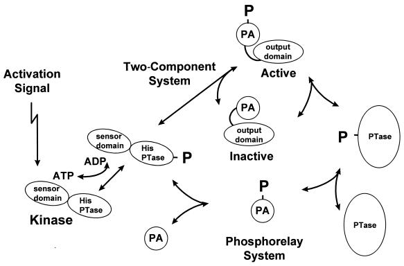
Schematic view of two-component and phosphorelay systems. Activation signals recognized by sensor domains of histidine kinases result in autophosphorylation of a histidine in the histidine phosphotransferase domain (His PTase). The phosphoryl group (P) is transferred directly to the phosphorylated aspartate domain (PA) of a response regulator in a two-component system, causing a conformational change that activates the output domain. In a phosphorelay, the phosphoryl group is transferred to a PA domain that serves as a substrate for a phosphotransferase whose role is to transfer the phosphoryl group to the PA domain of a response regulator. Note that all the steps are reversible in many systems, which may result in dephosphorylation in the absence of a signal.
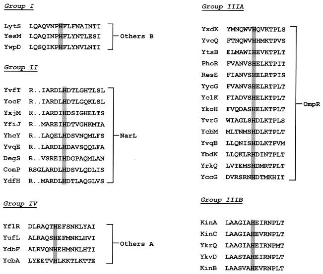
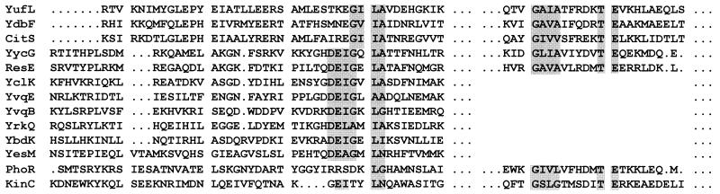
Classification of kinases by the sequence around the phosphorylated histidine. Kinases were sorted into classes on the basis of the sequence relationships of the residues on either side of the phosphorylated histidine. The classes were related to their response regulator based on the homology of the response regulator output domain to those of E. coli (19). Note the orphan kinases of group IIIB were related to NtrB of E. coli through the homology of the residues surrounding the histidine to NtrB, not through homologies to NtrC, a response regulator which does not exist in B. subtilis. Gaps introduced to maximize alignment are indicated by the dots.
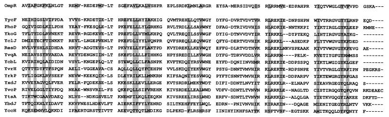
Relationships of response regulators to the output domain of OmpR. Amino acid sequences of response regulators were compared to the sequence of the output domain of OmpR of E. coli. The shaded residues are the residues making up the hydrophobic core of the domain (17). Gaps introduced to maximize alignment are indicated by the dashes.
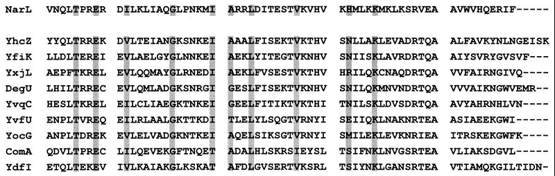
Relationships of response regulators to the output domain of NarL. Amino acid sequences of response regulators were compared to the sequence of the output domain of NarL of E. coli. The shaded residues are crucial for correct folding of the domain (2). Gaps introduced to maximize alignment are indicated by the dashes.
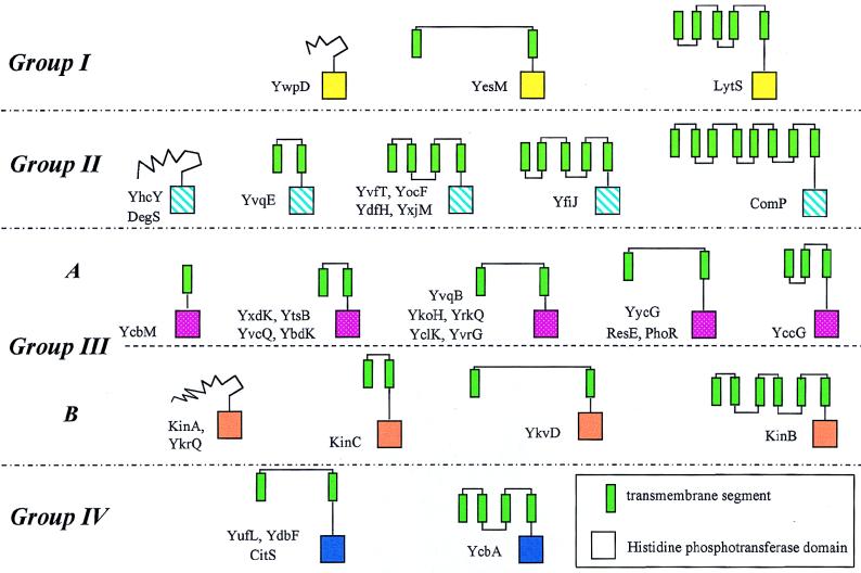
Schematic structures of the kinases. Groups were determined from the homology of the residues surrounding the phosphorylated histidine of the histidine phosphotransferase domain. Related domains are the same color, and green rectangles are likely transmembrane segments.
Similarities of sequences of cytoplasmic linkers. Sequences of linkers between the last transmembrane domain and the histidine motif of kinases that show homology are compared. The shaded residues define two motifs common to these linkers. Numerous partial homologies are not shaded for clarity. Gaps introduced to maximize alignment are indicated by the dots.
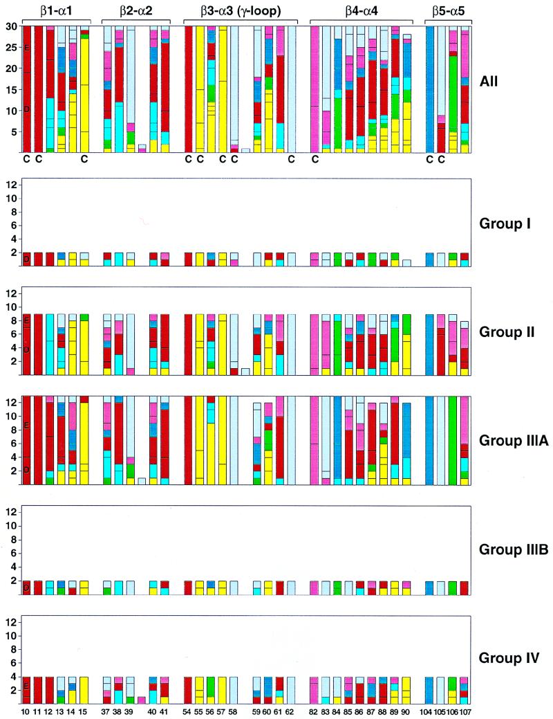
Sequence alignment of response regulator loop regions proximal to the site of phosphorylation for B. subtilis. The number of each response regulator residue type at loop positions spanning between secondary structure elements β-strand 1 to α-helix 1 (β1-α1), β2-α2, β3-α3, β4-α4, and β5-α5 are illustrated individually as members of groups I, II, IIIA, and IIIB, IV and collectively in the top bar graph for comparison. The residue type is coded by color: acidic in red (D and E), basic in dark blue (K and R), hydroxyl in rose (S and T), polar in light blue (H, N, and Q), hydrophobic in yellow (C, I, L, V, and M), aromatic in green (Y, F, and W), and structural in gray (A, G, and P). The numbering scheme is based on the Spo0F sequence, and conserved residues essential for phosphorylation are D10, D11, D54 (the phosphorylation site), T82, and K104 by this numbering. Residues previously described as conserved (C) by alignment of response regulators from all organisms (30) are denoted. Response regulator sequences (14) were aligned with Clustal W (10) and illustrated with the Excel 98 (Microsoft) and Adobe Illustrator programs (Adobe).
References
-
- Appleby J L, Parkinson J S, Bourret R B. Signal transduction via the multistep phosphorelay: not necessarily a road less traveled. Cell. 1996;86:845–848. - PubMed
-
- Baikalov I, Schroder I, Kaczor-Grzeskowiak M, Grzeskowiak K, Gunsalus R P, Dickerson R E. Structure of the Escherichia coli response regulator NarL. Biochemistry. 1996;35:11053–11061. - PubMed
-
- Birkey S M, Liu W, Zhang X, Duggan M F, Hulett F M. Pho signal transduction network reveals direct transcriptional regulation of one two-component system by another two-component regulator: Bacillus subtilis PhoP directly regulates production of ResD. Mol Microbiol. 1998;30:943–953. - PubMed
-
- Burbulys D, Trach K A, Hoch J A. The initiation of sporulation in Bacillus subtilis is controlled by a multicomponent phosphorelay. Cell. 1991;64:545–552. - PubMed
Publication types
MeSH terms
Substances
Grants and funding
LinkOut - more resources
Full Text Sources
Other Literature Sources
Molecular Biology Databases
