. 1999 Mar 30;96(7):3507-12.
doi: 10.1073/pnas.96.7.3507.

Design of highly specific cytotoxins by using trans-splicing ribozymes

Affiliations

Design of highly specific cytotoxins by using trans-splicing ribozymes

B G Ayre et al. Proc Natl Acad Sci U S A. .

Abstract

We have designed ribozymes based on a self-splicing group I intron that can trans-splice exon sequences into a chosen RNA target to create a functional chimeric mRNA and provide a highly specific trigger for gene expression. We have targeted ribozymes against the coat protein mRNA of a widespread plant pathogen, cucumber mosaic virus. The ribozymes were designed to trans-splice the coding sequence of the diphtheria toxin A chain in frame with the viral initiation codon of the target sequence. Diphtheria toxin A chain catalyzes the ADP ribosylation of elongation factor 2 and can cause the cessation of protein translation. In a Saccharomyces cerevisiae model system, ribozyme expression was shown to specifically inhibit the growth of cells expressing the virus mRNA. A point mutation at the target splice site alleviated this ribozyme-mediated toxicity. Increasing the extent of base pairing between the ribozyme and target dramatically increased specific expression of the cytotoxin and reduced illegitimate toxicity in vivo. Trans-splicing ribozymes may provide a new class of agents for engineering virus resistance and therapeutic cytotoxins.


Figures
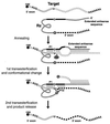
Figure 1
Figure 1
Ribozyme-mediated trans-splicing. Ribozyme and target RNAs are indicated schematically, with conserved sequences shown explicitly. In the first step, ribozyme and target RNAs base pair to form the P1 and extended antisense helices. Guanosine-mediated transesterification results in cleavage of the target RNA. The distal portion of helix P1 is displaced by sequences from the 3′ exon to form helix P10, which allows the second transesterification to proceed, and results in ligation of the two exons.
Figure 2
Figure 2
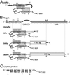
Design of ribozyme and target RNAs. (A) Schematic diagram of the cis-splicing ribozyme in pcisRz-DTA. The 5′-exon sequences are italic and uppercase, the 3′-exon sequences are nonitalic and uppercase, and intron sequences are lowercase. The IGS of the intron is represented in bold. The modified P1 helix sequences are shaded, and the modified P10 helix sequences are boxed. Watson-Crick base paring is indicated by |, and G:U base pairs are represented by dots. Arrows indicate the 5′- and 3′-splice sites. (B) Schematic diagram of the trans-splicing ribozymes. The target transcript is represented by the upper horizontal line with sequences around the splice site shown italicized. Trans-splicing ribozymes are represented below. Potential base pairing between the ribozyme and the target is indicated by |, and the length of the extended antisense sequences are indicated in rectangular boxes. The unpaired bulged region between the P1 helix and the extended antisense sequence is represented by the curved region in the target diagram. (C) Sequence of the expected ligated splice junction (indicated by arrows) and the encoded amino acids. Restriction endonuclease recognition sites: B, BamHI; X, XhoI; K, KpnI; S, SacI; P, PstI; N, NdeI; Bg, BglII. The figures are not drawn to scale.
Figure 3
Figure 3
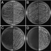
Targeted cell ablation with trans-splicing ribozymes and increased complementarity to the target RNA. Yeast strains expressing various ribozymes, pVT103-U (A), p9Rz-DTA (B), p54Rz-DTA (C), or p302Rz-DTA (D), were mated with strains containing the CMV target gene (pCMV-GFP, left) or a mutated version with a single nucleotide alteration at the 5′-splice site (pMut-GFP, right). The diploid progeny of the cross were plated onto selective media, and colony growth was recorded after 3 days.
Figure 4
Figure 4
Amplified products of in vivo cis- and trans-splicing. RT-PCR was used to amplify the RNA products of splicing from yeast cells expressing the indicated ribozyme and target genes. Bands migrating at a rate expected for DNA fragments 632 nt in length (indicated by an arrow) correspond to spliced products, and the major slower migrating bands in lanes 2 and 3 correspond to unspliced cis-acting ribozyme and inactive ΔP5abc derivative, respectively. A faint band corresponding to 632 nt was seen in lane 4.
Figure 5
Figure 5
Increasing the length of the antisense sequence reduces leaky expression of the toxin. The growth of haploid yeast cultures harboring indicated ribozyme was measured and normalized with respect to cultures containing the parental plasmid, pVT103-U (100%). The relative doubling times of each culture are represented by bars, white for active ribozymes and shaded for inactive ΔP5abc derivatives. The increased doubling times of cultures with active ribozymes relative to the corresponding ΔP5abc controls are shown as percentages on the diagram. Error bars correspond to the SEM from three experiments.

Similar articles

Cited by

References

    1. Cech T R. Annu Rev Biochem. 1990;59:543–568. - PubMed
    1. Galloway-Salvo J L, Coetzee T, Belfort M. J Mol Biol. 1990;211:537–549. - PubMed
    1. Sullenger B A, Cech T R. Nature (London) 1994;371:619–622. - PubMed
    1. Jones J T, Lee S W, Sullenger B A. Nat Med. 1996;2:643–648. - PubMed
    1. Sarver N, Cairns S. Nat Med. 1996;2:641–642. - PubMed

Publication types

MeSH terms

LinkOut - more resources