. 1999 Mar 30;96(7):3519-24.
doi: 10.1073/pnas.96.7.3519.

SMRTe, a silencing mediator for retinoid and thyroid hormone receptors-extended isoform that is more related to the nuclear receptor corepressor

Affiliations

SMRTe, a silencing mediator for retinoid and thyroid hormone receptors-extended isoform that is more related to the nuclear receptor corepressor

E J Park et al. Proc Natl Acad Sci U S A. .

Abstract

SMRT (silencing mediator for retinoid and thyroid hormone receptors) and N-CoR (nuclear receptor copressor) mediate transcriptional repression of important regulators that are involved in many signaling pathways. SMRT and N-CoR are related proteins that form complexes with mSin3A/B and histone deacetylases to induce local chromatin condensation and transcriptional repression. However, SMRT is substantially smaller than N-CoR, lacking an N-terminal domain of approximately 1,000 aa that are present in N-CoR. Here, we report the identification of SMRT-extended (SMRTe), which contains an N-terminal sequence that shows striking similarity with N-CoR. As in N-CoR, this SMRTe-N-terminal domain also represses basal transcription. We find that SMRTe expression is regulated during cell cycle progression and SMRTe transcripts are present in many embryonic tissues. These data redefine a structurally and functionally more related nuclear receptor corepressor family and suggest an additional role for SMRTe in the regulation of cycle-specific gene expression in diverse signaling pathways.


Figures

Figure 1
Figure 1
Detection of endogenous SMRT proteins. HeLa nuclear extract, together with in vitro-translated [35S]methionine-labeled N-CoR and C-SMRT, were separated on a SDS/PAGE. The N-CoR and C-SMRT polypeptides were detected by autoradiography (Left). An identical gel was processed for Western blotting using an affinity-purified rabbit anti-C-SMRT polyclonal antibody and detected by 5-bromo-4-chloro-3-indolyl phosphate (BCIP)/nitroblue tetrazolium (NBT) color reaction (Center). One major polypeptide similar to the size of N-CoR (270 kDa) was detected in the HeLa nuclear extract, in addition to two minor bands of 180 and 80 kDa, respectively (arrows). The anti-SMRT antibody does not crossreact with N-CoR. The same HeLa nuclear extract also was processed for Western blotting using anti-C-SMRT antibody but developed by ECL+ reaction (Right). The three specific SMRT polypeptides and two nonspecific bands (open arrowheads) below 80 kDa were indicated.
Figure 2
Figure 2
Sequences and domain comparison of mouse and human SMRTe and N-CoR. (A) The amino acid sequence of human (h) SMRTe is shown in the upper strand, and the mouse (m) SMRTe sequence is in the bottom strand. Identical amino acids between human and mouse SMRTe are indicated by hyphens. Dots are gaps introduced during the alignment. The COOH-terminal tail of the mSMRTeΔC (ΔC), the starting amino acids of the previously identified SMRT and TRAC1 also are indicated. (B) Domain comparison between SMRTe and N-CoR. The black bars indicate areas of high homology. Special domains are indicated in gray with labels (AB, acidic-basic domain; S1–4, the SIT repeated motifs, ref. ; KGH, the KGH repeated motifs, ref. ; SG, the serine/glycine-rich region; and SNC). The SMRTe repression domains (SRD), the N-CoR repression domains (NRD), and the nuclear receptor interacting domains (RID) are also shown. Domains involved in interactions with other proteins are also indicated. The numbering of residues is based on mouse N-CoR and human SMRTe sequence. (C) Sequence comparison of the SNC domains of human (h) and mouse (m) SMRTe (S) and N-CoR (N). The identical residues are shown in black and the conserved residues are shown in gray. The amphipathic helix and the hydrophobic heptad repeats are indicated by a black line and stars, respectively. The amino acid residues are shown on the left. (D) Comparison of SANT-A and SANT-B domains. Identical amino acids are shown in black background and the conserved residues are in gray. The Myb DNA binding domain signature sequences and the three helices (h) are also indicated in between the SANT-A and SANT-B motifs.
Figure 3
Figure 3
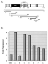
Transcriptional repression by the N-terminal domain of SMRTe. (A) Schematic of the Gal4 DBD fusion constructs used in this experiment. The domains are as described in Fig. 2B. The numbers indicate amino acid residues. The seven different SMRTe N-terminal fragments (A to G) are fused into Gal4 DBD and their effects on reporter gene expression are shown in B. (B) Transcriptional repression by Gal4 DBD-SMRTe fusions. The fold repression of each construct was determined by average relative luciferase activity using Gal4 DBD as a standard in a triplicate experiment.
Figure 4
Figure 4
Cell cycle-dependent expression of SMRTe. (A) Immunofluorescence staining of endogenous SMRTe in HeLa cells. The nucleus stained by 4′,6-diamidino-2-phenylindole (DAPI) dihydrochloride hydrate (Upper) and the SMRTe signal (Lower) is shown. Note that SMRTe is distributed as fine granules in the nucleus and is excluded from the nucleoli. (B) Immunostaining of SMRTe in an unsynchronized population of A549 cells. Strong SMRTe signals were detected in a subset of A549 cells. (C) Western blotting for SMRTe in A549 cells at different time points after release from mitosis. The 270-kDa SMRTe signal (Upper) and a nonspecific band (Lower) are shown.
Figure 5
Figure 5
SMRTe transcripts in mouse embryos. SMRTe transcripts were detected by in situ hybridization to thin sections of (A) E9.0 days postconception, (B) E11.5, and (C) E13.5 using DIG-labeled antisense riboprobe. c1 and c2 show enlargement of areas in the cartilage and lung at E13.5 indicated by rectangles in C. (D) DIG-labeled sense probe shows the background signal. b, brain; ba, bronchial arch; br, bronchus; c, cartilage; cp, cerebellar plate; h, heart; lm, limb; lu, lung; lv, liver; nt, neural tube; pc, perichondrium; sc, sclerotome; vb, vertebra body.

Similar articles

Cited by

References

    1. Johnson A D. Cell. 1995;81:655–658. - PubMed
    1. Hanna-Rose W, Hansen U. Trends Genet. 1996;12:229–234. - PubMed
    1. DePinho R A. Nature (London) 1998;391:533. , 535–536. - PubMed
    1. Baniahmad A, Kohne A C, Renkawitz R. Cell. 1992;11:1015–1023. - PMC - PubMed
    1. Chen J D, Evans R M. Nature (London) 1995;377:454–457. - PubMed

Publication types

MeSH terms

Associated data