. 1999 Mar 30;96(7):3556-61.
doi: 10.1073/pnas.96.7.3556.

The role of threonine 37 in flavin reactivity of the old yellow enzyme

Affiliations

The role of threonine 37 in flavin reactivity of the old yellow enzyme

D Xu et al. Proc Natl Acad Sci U S A. .

Abstract

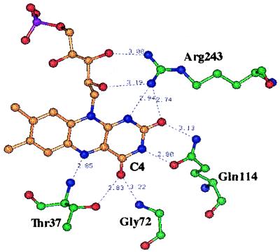
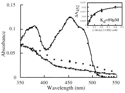
Threonine 37 is conserved among all the members of the old yellow enzyme (OYE) family. The hydroxyl group of this residue forms a hydrogen bond with the C-4 oxygen atom of the FMN reaction center of the enzyme [Fox, K. M. & Karplus, P. A. (1994) Structure 2, 1089-1105]. The position of Thr-37 and its interaction with flavin allow for speculations about its role in enzyme activity. This residue was mutated to alanine and the mutant enzyme was studied and compared with the wild-type OYE1 to evaluate its mechanistic function. The mutation has different effects on the two separate half-reactions of the enzyme. The mutant enzyme has enhanced activity in the oxidative half-reaction but the reductive half-reaction is slowed down by more than one order of magnitude. The peaks of the absorption spectra for enzyme bound with phenolic compounds are shifted toward shorter wavelengths than those of wild-type OYE1, consistent with its lower redox potential. It is suggested that Thr-37 in the wild-type OYE1 increases the redox potential of the enzyme by stabilizing the negative charge of the reduced flavin through hydrogen bonding with it.


Figures

Figure 1
Figure 1
The FMN binding site of OYE1. Hydrogen bonds are shown as dashed lines and the distance between the atom attached to the donor hydrogen and the acceptor is shown in Å (Protein Data Bank ID code 1OYB). In this projection, the viewer is looking down on the si-face of the flavin. In this figure, H-atoms have been eliminated; N-atoms are displayed in blue, O-atoms in red, flavin C-atoms in orange, the P-atom in purple, and amino acid C-atoms in green.
Figure 2
Figure 2
Primer selection for mutagenesis. In the mutagenic primer, the underlined codon includes the italicized substitution that encodes the Thr-37 to Ala mutation. The solid arrows describe the endpoints for the PCR1 reaction product; the dashed arrows describe the endpoints for the PCR2 reaction. The mutagenesis primer contains the desired Thr-37 to Ala mutation with a TGG → CGG substitution in the Thr-37 codon that introduces an analytically useful MscI restriction site (ACC/GGT).
Figure 3
Figure 3
Spectral comparison of oxidized and p-chlorophenol-bound wild-type OYE1 and the T37A mutant. The spectral data are scaled so the wild-type absorbance 462 nm agrees with the T37A absorbance at 452 nm, corresponding to 15 μM wild-type enzyme. The solid lines and open symbols are for the T37A mutant enzyme; the dashed lines and closed symbols represent the wild-type OYE1 spectra. The spectra with long wavelength absorbance marked by squares are the result of addition of saturating (500 μM) p-chlorophenol to the enzyme.
Figure 4
Figure 4
Redox potential measurement of OYE1-T37A. (Upper) Oxidized enzyme (35 μM, 1.2 ml) was titrated in an anaerobic cuvette with NADPH (closed circles) or NADH (open circles). The redox potential is acquired as the value of log ([Ox]/[Red]) when log ([NAD(P)+]/[NAD(P)H]) = 0. (Lower) Correlation of the absorbance peak positions of the charge-transfer complex formed by OYE1 and p-chlorophenol and the redox potential of the coenzyme. The open circles represent the results of wild-type enzyme with flavin analogues bound (refs. 22, 23). The result with T37A is shown as the closed circle.
Figure 5
Figure 5
Reductive half-reaction of T37A-OYE1. Oxidized enzyme (final concentration 15 μM) was reacted anaerobically with NADPH (final concentration 100 μM) in the stopped-flow apparatus. The absorbance change was recorded at 450 nm (dashed line) and 550 nm (solid line).
Figure 6
Figure 6
The Michaelis complex intermediate of 3-methyl-2-cyclohexenone and reduced OYE1-T37A. The reduced enzyme was mixed with the substrate (final concentrations 12 μM and 200 μM, respectively), and the dead time absorbances (3 ms) at various wavelengths (open circles) are shown and proposed to be the spectrum of the Michaelis complex. The spectra of the reduced (open squares) and oxidized (open triangles) mutant enzyme, scaled to the same concentration, are also shown. (Insert) The absorbance change at 452 nm during the dead time of the stopped-flow experiments is plotted against the final concentration of the 3-methyl-2-cyclohexenone.

Similar articles

Cited by

References

    1. Warburg O, Christian W. Biochem Z. 1933;266:377–411.
    1. Theorell H. Biochem Z. 1935;275:344–346.
    1. Stott K, Saito K, Thiele D J, Massey V. J Biol Chem. 1993;268:6097–6106. - PubMed
    1. Abramovitz A S, Massey V. J Biol Chem. 1976;251:5327–5336. - PubMed
    1. Massey V, Schopfer L M. J Biol Chem. 1986;261:1215–1222. - PubMed

Publication types

LinkOut - more resources