. 1999 Mar 30;96(7):3562-7.
doi: 10.1073/pnas.96.7.3562.

Combinatorial protein engineering by incremental truncation

Affiliations

Combinatorial protein engineering by incremental truncation

M Ostermeier et al. Proc Natl Acad Sci U S A. .

Abstract

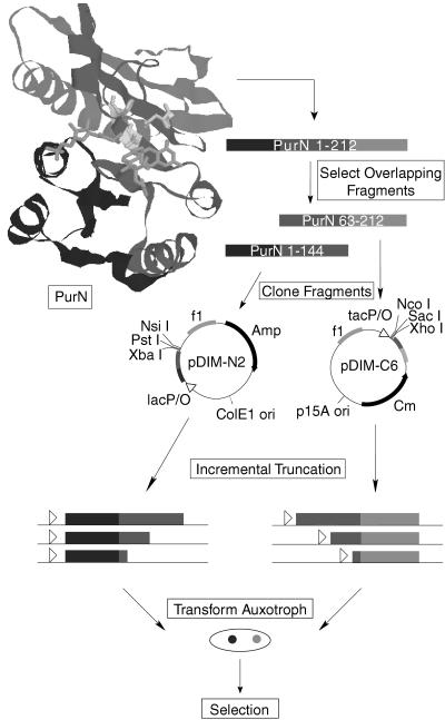
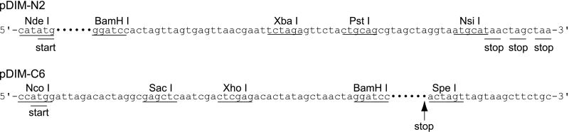
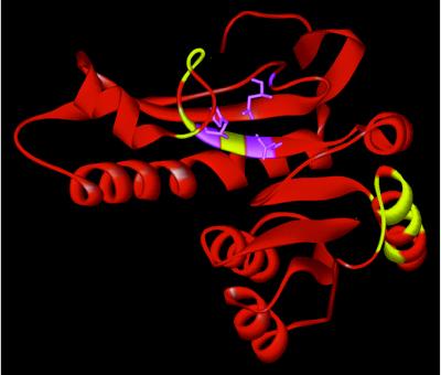
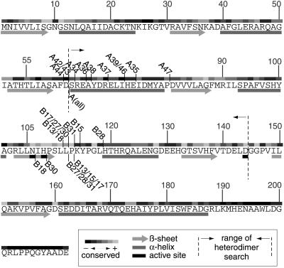
We have developed a combinatorial approach, using incremental truncation libraries of overlapping N- and C-terminal gene fragments, that examines all possible bisection points within a given region of an enzyme that will allow the conversion of a monomeric enzyme into its functional heterodimer. This general method for enzyme bisection will have broad applications in the engineering of new catalytic functions through domain swapping and chemical synthesis of modified peptide fragments and in the study of enzyme evolution and protein folding. We have tested this methodology on Escherichia coli glycinamide ribonucleotide formyltransferase (PurN) and, by genetic selection, identified PurN heterodimers capable of glycinamide ribonucleotide transformylation. Two were chosen for physical characterization and were found to be comparable to the wild-type PurN monomer in terms of stability to denaturation, activity, and binding of substrate and cofactor. Sequence analysis of 18 randomly chosen, active PurN heterodimers revealed that the breakpoints primarily clustered in loops near the surface of the enzyme, that the breaks could result in the deletion of highly conserved residues and, most surprisingly, that the active site could be bisected.


Figures

Figure 1
Figure 1
Experimental overview. Based on the structure of PurN (16), the purN gene was divided into nonactive overlapping fragments. The N-terminus fragment (purN[1–144]) consists of the DNA coding for residues 1–144 and the C-terminus fragment (purN[63–212]) consists of the DNA coding for residues 63–212. By experimental design, the cut-point search is limited to the region of overlap between the two fragments (63–144). The two fragments were cloned into vectors pDIM-N2 and pDIM-C6 designed for incremental truncation. Libraries of incremental truncations from the C terminus of the purN[1–144] gene fragment and from the N terminus of the purN[63–212] gene fragment were created by using Exo III digestion at low temperatures. These incremental truncation libraries then were transformed together into an auxotrophic E. coli strain unable to grow on minimal media lacking purines unless GAR transformylase activity is supplied in trans. Active PurN heterodimers then were selected by plating the crossed library onto selective minimal plates. Colonies that grew contained an active PurN enzyme.
Figure 2
Figure 2
Vectors for incremental truncation. The •••••• sequence corresponds to the sequence of the gene fragment to be truncated. The start and stop codons for the incremental truncation libraries are shown. The stop codon for pDIM-C6 is at the 3′ end of the gene fragment to be truncated.
Figure 3
Figure 3
Cut-points mapped onto the crystal structure of PurN (16). The new N and C termini for all the functional PurN heterodimers are indicated in yellow. Active site residues N106, H108, and D144 are indicated in violet. The original C terminus of the wild-type monomer is at the top.
Figure 4
Figure 4
Cut-points relative to secondary structure and conserved residues. The cut-points of all positives sequenced are shown relative to the amino acid sequence of PurN. If the number of the positive is shown above the sequence, it indicates the end of an N-terminal fragment. If the number of the positive is shown below the sequence, it indicates the beginning of the C-terminal fragment. For the A series of positives, all C-terminal fragments start at the same residue indicated by A(all). The degree of conservation of each amino acid is indicated above the sequence and was determined by alignment of 16 GAR transformylases from E. coli to human by using megalign (DNASTAR, Madison, WI). Secondary structure (16) is indicated below the sequence.

Similar articles

Cited by

References

    1. Kato I, Anfinsen C B. J Biol Chem. 1969;244:1004–1007. - PubMed
    1. Ullmann A, Jacob F, Monod J. J Mol Biol. 1967;24:339–343. - PubMed
    1. Bertolaet B L, Knowles J R. Biochemistry. 1995;34:5736–5743. - PubMed
    1. Ladurner A G, Itzhaki L S, Gray G P, Fersht A R. J Mol Biol. 1997;273:317–329. - PubMed
    1. Tasayco M L, Carey J. Science. 1992;255:594–597. - PubMed

Publication types

MeSH terms

Substances

LinkOut - more resources