. 1999 Mar 30;96(7):3572-7.
doi: 10.1073/pnas.96.7.3572.

Crystal structure of human p32, a doughnut-shaped acidic mitochondrial matrix protein

Affiliations

Crystal structure of human p32, a doughnut-shaped acidic mitochondrial matrix protein

J Jiang et al. Proc Natl Acad Sci U S A. .

Abstract

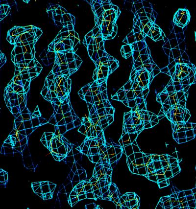
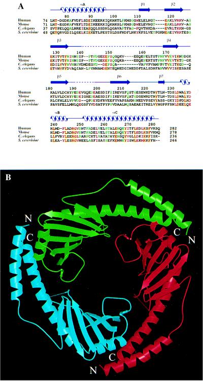
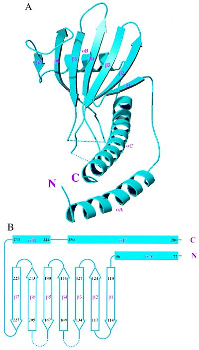
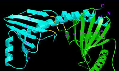
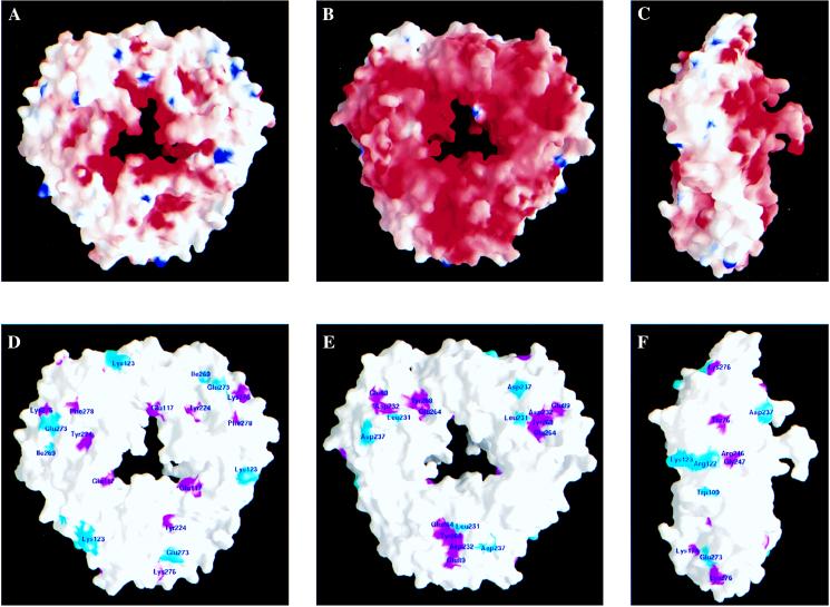
Human p32 (also known as SF2-associated p32, p32/TAP, and gC1qR) is a conserved eukaryotic protein that localizes predominantly in the mitochondrial matrix. It is thought to be involved in mitochondrial oxidative phosphorylation and in nucleus-mitochondrion interactions. We report the crystal structure of p32 determined at 2.25 A resolution. The structure reveals that p32 adopts a novel fold with seven consecutive antiparallel beta-strands flanked by one N-terminal and two C-terminal alpha-helices. Three monomers form a doughnut-shaped quaternary structure with an unusually asymmetric charge distribution on the surface. The implications of the structure on previously proposed functions of p32 are discussed and new specific functional properties are suggested.


Figures

Figure 1
Figure 1
MAD-phased electron density map. A portion of the MAD-phased electron density covering the central β-strands is shown with a refined model superimposed onto the map. The electron density was computed to 2.5 Å resolution and contoured at 1.2 σ level.
Figure 2
Figure 2
Overall structure of p32. (A) Structure-based alignment of p32 sequences from human (PID g338043), mouse (PID g743485), Caenorhabditis elegans (PID g3334445) and S. cerevisiae (PID g557799). Only the sequences corresponding to the mature human protein are shown. Positions with four identical amino acids are shown in red letters over yellow background; positions with four similar amino acids are shown in plain red letters; positions with three identities are shown in green letters. Related expressed sequence tag fragments or genomic sequences can also be found in Drosophila (NID g3112239), zebrafish (NID g2446858), and Arabidopsis (PID g3334441). PID and NID are protein and nucleotide identification numbers in GenBank. (B) Ribbon representation of the p32 trimer, looking down the noncrystallographic three-fold axis. The dotted lines show disordered segments in the structure. The three monomers that make up the trimer are colored red, green and cyan, and are referred to as subunits A, B, and C, respectively. The same color codes are used in other figures.
Figure 3
Figure 3
Folding topology of p32. (A) Ribbon representation of a p32 monomer. The secondary structure elements are labeled. (B) Schematic drawing of the p32 topology.
Figure 4
Figure 4
Subunit interface between p32 monomers. The interactions between monomers B (green) and C (cyan) within the p32 trimer are shown, as viewed from the inside of the channel. The portions of the two loops in subunit C and one loop in subunit B that interact with each other (see text) are indicated in yellow. In addition, extensive intermolecular interactions involve contacts between αC and αA of monomer C with the β-sheet and αB of monomer B, respectively, as described in the text.
Figure 5
Figure 5
Surface distribution of charged and phylogenetically conserved amino acids. (A) Charge distribution on the protein surface, viewed from the same direction as in Fig. 1B. This side is referred to as the positive side. Positively and negatively charged electric potential are indicated in blue and red, respectively. The surface was calculated by using a probe radius of 1.4 Å, and the potential is displayed at the scale of −20 kBT to +20 kBT, where kB is the Boltzmann constant. (B) A significantly higher number of negatively charged residues is present on the opposite side of the protein surface. This side is referred to as the negative side. (C) A side view of the protein surface shows the asymmetry of charge distribution on the two sides of the protein surface. (D) Distribution of conserved amino acids on the positive side of the protein surface. Magenta indicates identical residues, and cyan indicates similar residues. They are shown in Fig. 2A as red letters over yellow background and plain red letters, respectively. (E) Several conserved and spatially clustered residues form a shallow pocket on the negative side of the protein surface. (F) Distribution of conserved amino acids on the outer perimeter of the protein surface.

References

    1. Krainer A R, Mayeda A, Kozak D, Binns G. Cell. 1991;66:383–394. - PubMed
    1. Desai K, Loewenstein P M, Green M. Proc Natl Acad Sci USA. 1991;88:8875–8879. - PMC - PubMed
    1. Yu L, Zhang Z, Loewenstein P M, Desai K, Tang Q, Mao D, Symington J S, Green M. J Virol. 1995;69:3007–3016. - PMC - PubMed
    1. Fridell R A, Harding L S, Bogerd H P, Cullen B R. Virology. 1995;209:347–357. - PubMed
    1. Luo Y, Yu H, Peterlin B M. J Virol. 1994;68:3850–3856. - PMC - PubMed

Publication types

MeSH terms

Associated data