. 1999 Mar 30;96(7):3658-63.
doi: 10.1073/pnas.96.7.3658.

Molecular characteristics of fibroblast growth factor-fibroblast growth factor receptor-heparin-like glycosaminoglycan complex

Affiliations

Molecular characteristics of fibroblast growth factor-fibroblast growth factor receptor-heparin-like glycosaminoglycan complex

G Venkataraman et al. Proc Natl Acad Sci U S A. .

Abstract

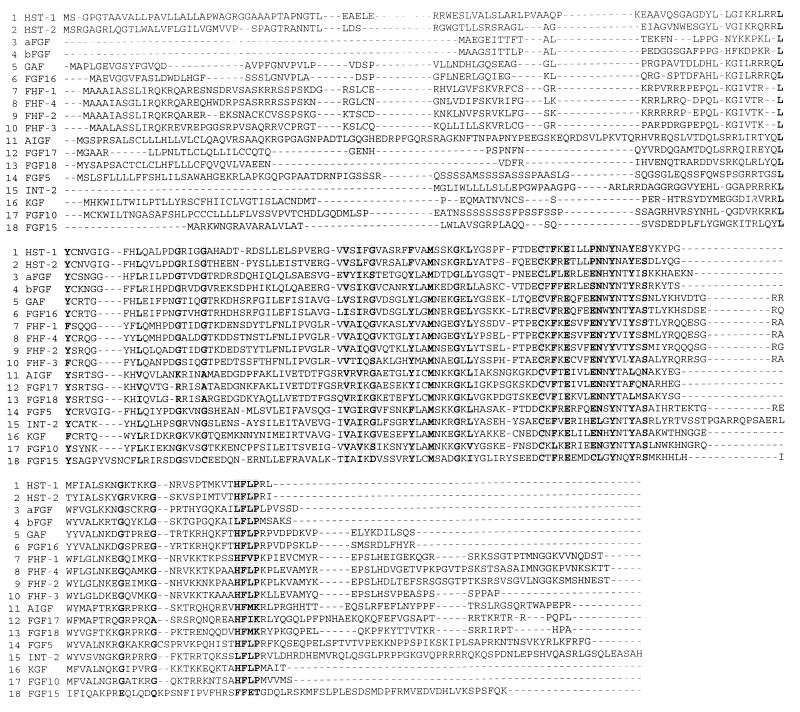
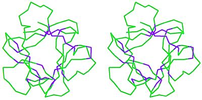
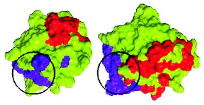
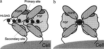
Fibroblast growth factor (FGF) family plays key roles in development, wound healing, and angiogenesis. Understanding of the molecular nature of interactions of FGFs with their receptors (FGFRs) has been seriously limited by the absence of structural information on FGFR or FGF-FGFR complex. In this study, based on an exhaustive analysis of the primary sequences of the FGF family, we determined that the residues that constitute the primary receptor-binding site of FGF-2 are conserved throughout the FGF family, whereas those of the secondary receptor binding site of FGF-2 are not. We propose that the FGF-FGFR interaction mediated by the 'conserved' primary site interactions is likely to be similar if not identical for the entire FGF family, whereas the 'variable' secondary sites, on both FGF as well as FGFR mediates specificity of a given FGF to a given FGFR isoform. Furthermore, as the pro-inflammatory cytokine interleukin 1 (IL-1) and FGF-2 share the same structural scaffold, we find that the spatial orientation of the primary receptor-binding site of FGF-2 coincides structurally with the IL-1beta receptor-binding site when the two molecules are superimposed. The structural similarities between the IL-1 and the FGF system provided a framework to elucidate molecular principles of FGF-FGFR interactions. In the FGF-FGFR model proposed here, the two domains of a single FGFR wrap around a single FGF-2 molecule such that one domain of FGFR binds to the primary receptor-binding site of the FGF molecule, while the second domain of the same FGFR binds to the secondary receptor-binding site of the same FGF molecule. Finally, the proposed model is able to accommodate not only heparin-like glycosaminoglycan (HLGAG) interactions with FGF and FGFR but also FGF dimerization or oligomerization mediated by HLGAG.


Figures

Figure 1
Figure 1
Multiple sequence alignment of FGF-1 to FGF-18 by the clustalw program (22). Boxes mark regions of highly conserved amino acid residues as described in Methods. FGF-1 to FGF-9 and FHF-11 to FGF-14 were obtained from the SwissProt database. FGF-10 and FGF-15 to FGF-18 were obtained from the GenBank database. The accession numbers for the FGFs are as follows: FGF-1 (acidic FGF, aFGF), P05230; FGF-2 (basic FGF, bFGF), P09038; FGF-3 (INT-2, protooncogene protein precursor), P11487; FGF-4 (KS3 or heparin secretory transforming protein 1, HST-1), P08620; FGF-5, P12034; FGF-6 (heparin secretory transforming protein 2, HST-2), P10767; FGF-7 (keratinocyte growth factor, KGF), P21781; FGF-8 (androgen-induced growth factor, AIGF), P55075; FGF-9 (glia-activating factor, GAF), P54130; FGF-10, 2911146; FGF-11 (FGF homologous factor 3, FHF-3), Q92914; FGF-12 (FGF homologous factor 1, FHF-1), Q92912; FGF-13 (FGF homologous factor 2, FHF-2), Q92913; FGF-14 (FGF homologous factor 4, FHF-4), Q92915; FGF-15, 2257959; FGF-16, 2911170; FGF-17, 3041790; and FGF-18, 3355904.
Figure 2
Figure 2
Stereo representation of the Cα trace of the FGF-2 molecule. The conserved amino acids were mapped onto the FGF-2 crystal structure (Protein Data Bank ID: 1fga). The conserved amino acids shown in Fig. 1 are colored purple, while the rest of the Cα atoms are colored green. Most of the conserved amino acids fall within the core region of the FGF-2 structure, whereas few of the amino acids are close to the FGF-2 surface.
Figure 3
Figure 3
The conserved solvent-accessible residues on the surface of the FGF-2 structure. These residues form a van der Waals surface patch. A Connolly surface rendering of the FGF-2 generated by using Insight II with the solvent-accessible conserved amino acid residues colored in purple is shown. This site coincides with the primary receptor binding site identified on the basis of site-directed mutagenesis studies (33).
Figure 4
Figure 4
Primary and secondary receptor binding sites of FGF-2. The binding sites are identified on the basis of site-directed mutagenesis studies (–35). (a) On the left is the Connolly surface rendering of the FGF-2 molecule with the residues corresponding to the primary site in purple and those corresponding to the secondary site in red. Notice that the secondary site (red) is not visible in this view as it is on the diametrically opposite end of the molecule compared with the primary site. (b) Same molecule as in a but rotated about a vertical axis in the plane of the paper by 180° so as to view the molecule from the other side.
Figure 5
Figure 5
Comparison of the spatial orientation of the primary (purple) and secondary (red) receptor-binding sites of FGF-2 and the primary (purple) and secondary (red) receptor-binding sites of IL-1β (31). The Cα traces of the FGF-2 molecule and the IL-1β molecule were superimposed. The FGF-2 molecule was then translated horizontally for comparison. The primary binding site (highlighted with a black circle) of FGF-2 forms a part of the primary binding site of IL-1β [identified on the basis of the interaction with receptor domains I and II in the cocrystal structure (31)]. The relative spatial orientations of the two primary binding sites are similar (when superimposed they are at the bottom left of the molecules). The number of residues constituting the IL-1β primary binding site is much higher than in FGF-2. Although the three-dimensional orientations of the set of noncontiguous amino acids that confer receptor binding are very similar in the two proteins, there is no one-to-one correspondence in the amino acid composition at the receptor-binding sites. Note also that the secondary binding sites (red), which are believed to provide functional specificity, are spatially located differently in FGF-2 and IL-1β.
Figure 6
Figure 6
(a) Molecular model of the FGF–FGFR complex. FGF-2 molecule (Connolly surface rendering, colored in green) interacts with the Ig II and Ig III domains of the receptors. The Ig III domain of the receptor (bottom of the figure) interacts with the secondary binding site of FGF-2 (red) whereas the Ig II domain of FGFR interacts with the primary binding site of FGF-2 (purple). The Ig I domain of the receptor is not shown in this model. Notice that the linker between Ig II and Ig III is long and flexible to position the two Ig domains at diametrically opposite ends of the FGF-2 molecules. Extension of this model to the other members of the FGF family would suggest that the interaction at the primary site (purple and Ig II) would remain the same, whereas the interaction of the secondary site (Ig III and red) can vary to topologically other regions of the molecule. (b) A model of FGF-2–HLGAG–FGFR interaction. The HLGAG-binding site of FGF-2 is sandwiched between the primary and the secondary receptor-binding sites. HLGAG can bind to FGF-2 such that it interacts with both the receptor domains and the linker between the domains.
Figure 7
Figure 7
(a) Schematic representation of the receptor clustering and oligomerization resulting from a side-by-side oligomerization of FGF as in a “beads-on-a-string” model. (b) Schematic representation of the receptor dimerization induced by a FGF-1–HLGAG dimer as observed in the cocrystal structure of FGF-1–decasaccharide (41). This model is schematically similar to the model suggested by DiGabrielle et al. (41).

Similar articles

Cited by

References

    1. Conrad E. Heparin Binding Proteins. New York: Academic; 1998. pp. 301–348.
    1. Basilico C, Moscatelli D. Adv Cancer Res. 1992;59:115–165. - PubMed
    1. Sasisekharan R, Ernst S, Venkataraman G. Angiogenesis. 1997;1:45–54. - PubMed
    1. Friesel R B, Maciag T. FASEB J. 1995;9:919–925. - PubMed
    1. Smallwood P M, Munozsanjuan I, Tong P, Macke J P, Hendry S H C, Gilbert D J, Copeland N G, Jenkins N A, Nathans J. Proc Natl Acad Sci USA. 1996;93:9850–9857. - PMC - PubMed

Publication types

MeSH terms

LinkOut - more resources