. 1999 Mar 30;96(7):3688-93.
doi: 10.1073/pnas.96.7.3688.

In situ atomic force microscopy study of Alzheimer's beta-amyloid peptide on different substrates: new insights into mechanism of beta-sheet formation

Affiliations

In situ atomic force microscopy study of Alzheimer's beta-amyloid peptide on different substrates: new insights into mechanism of beta-sheet formation

T Kowalewski et al. Proc Natl Acad Sci U S A. .

Abstract

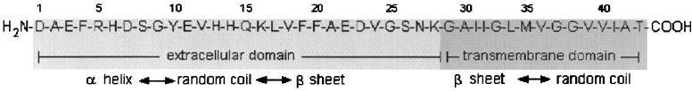
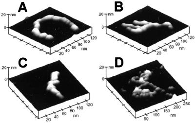
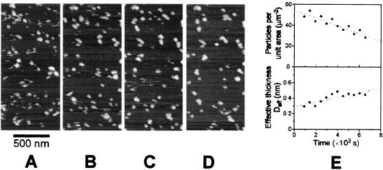
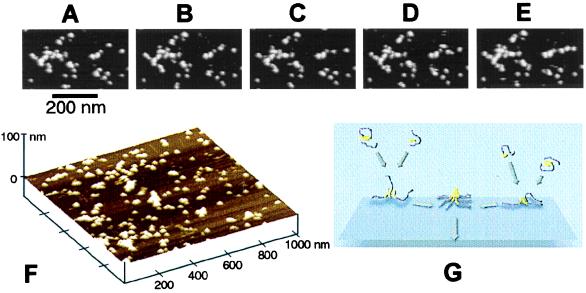
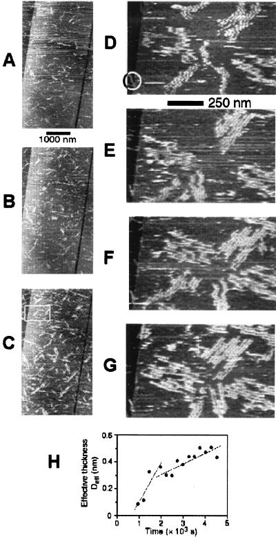
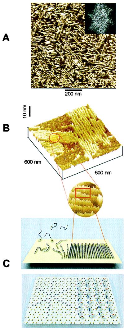
We have applied in situ atomic force microscopy to directly observe the aggregation of Alzheimer's beta-amyloid peptide (Abeta) in contact with two model solid surfaces: hydrophilic mica and hydrophobic graphite. The time course of aggregation was followed by continuous imaging of surfaces remaining in contact with 10-500 microM solutions of Abeta in PBS (pH 7.4). Visualization of fragile nanoscale aggregates of Abeta was made possible by the application of a tapping mode of imaging, which minimizes the lateral forces between the probe tip and the sample. The size and the shape of Abeta aggregates, as well as the kinetics of their formation, exhibited pronounced dependence on the physicochemical nature of the surface. On hydrophilic mica, Abeta formed particulate, pseudomicellar aggregates, which at higher Abeta concentration had the tendency to form linear assemblies, reminiscent of protofibrillar species described recently in the literature. In contrast, on hydrophobic graphite Abeta formed uniform, elongated sheets. The dimensions of those sheets were consistent with the dimensions of beta-sheets with extended peptide chains perpendicular to the long axis of the aggregate. The sheets of Abeta were oriented along three directions at 120 degrees to each other, resembling the crystallographic symmetry of a graphite surface. Such substrate-templated self-assembly may be the distinguishing feature of beta-sheets in comparison with alpha-helices. These studies show that in situ atomic force microscopy enables direct assessment of amyloid aggregation in physiological fluids and suggest that Abeta fibril formation may be driven by interactions at the interface of aqueous solutions and hydrophobic substrates, as occurs in membranes and lipoprotein particles in vivo.


Figures

Figure 1
Figure 1
The sequence of Aβ peptide.
Figure 2
Figure 2
Ex situ tapping mode AFM images of individual protofibrillar aggregates of Aβ(1–42) deposited on mica. The height in these and all subsequent images was color coded, with lighter tones corresponding to taller features.
Figure 3
Figure 3
Aggregation of Aβ on mica. (AD) Tapping mode AFM images acquired in situ in a 10 μM solution of Aβ in PBS. The images correspond to the same area of mica and were digitally zoomed-in from 3- × 3-μm datasets. The images were aligned with respect to the features located in the upper left corner as well as with respect to additional reference points located outside the zoomed-in field of view. Nanoparticulate aggregates of Aβ appeared on the surface almost immediately after immersion in solution. They behaved as nanodroplets of a substance poorly wetting the substrate, exhibiting marked lateral mobility and a tendency to coalesce. The time interval between the images was 2,048 s. (E) Results of quantitative analysis of aggregation. As a function of time the number of particles per unit area decreased (top), whereas the volume of material deposited per unit area, i.e., the effective thickness Deff increased (bottom). Such behavior indicates the coalescence of nanodroplets. The analysis was performed on a 4.5-μm2 area of original 3- × 3-μm images.
Figure 4
Figure 4
In situ tapping mode AFM images of aggregation of Aβ in contact with mica at high peptide concentration (500 μM). All images were digitally zoomed in from a 3- × 3-μm scan. (AE) Time course of the process of assembly of globular aggregates into a linear protofibril located on the right side of an image. Notice that the branched protofibrillar assembly located on the left side remained mostly static over the same period. The time interval between the images was 256 s. (F) Three-dimensional rendering of the surface of mica covered with globular and protofibrillar aggregates of Aβ. (G) Schematic of the proposed mechanism of the early stages of formation of pseudomicellar aggregates of an amphipathic peptide on a hydrophilic substrate. (The hydrophilic and hydrophobic portions of peptide chains are colored, respectively, in purple and in yellow.)
Figure 5
Figure 5
Aggregation of Aβ on graphite observed with in situ tapping mode AFM in a 10 μM solution of Aβ(1–42) in PBS. (AC) The first phase of aggregation i.e., rapid appearance of highly elongated, narrow, sheet-like aggregates. The images were digitally zoomed in from 10- × 10-μm consecutive original scans; the time interval between the images is equal to 256 s. (DG) The second phase of aggregation i.e., formation of parallel assemblies of sheets. The images correspond to the area marked with a rectangle in C. The images were digitally zoomed in from 4- × 4-μm original scans and were aligned with respect to the feature marked with a circle in D; the time interval between the images is equal to 512 s. (H) Quantitative analysis of aggregation. The effective thickness of adsorbed material, Deff, is shown as a function of time. Notice the presence of an incubation time and two regimes of growth.
Figure 6
Figure 6
(A) An example of preferential orientation of sheet-like aggregates of Aβ and their assemblies forming on graphite. The orientation of aggregates along the three directions at 120° to one another is reflected in the characteristic 6-fold symmetry of the two-dimensional Fourier transform of the image (Inset). The average lateral spacing between the sheets, determined from the radial position of spots in Fourier transform patterns of images from different experiments, was equal to 18.8 ± 1.8 nm. (B) Higher magnification view of two assemblies of Aβ aggregates on graphite (top) and a schematic model illustrating the orientation of peptide chains in the aggregates based on their dimensions (bottom). The height of the aggregates above the graphite surface, measured from their profiles ranged from 1.0 to 1.2 nm. Dimensions of Aβ aggregates on graphite provide a strong indication that they have a β-sheet character, with peptide chains perpendicular to the aggregate long axis. (C) The properly scaled model of peptide backbones in an antiparallel β-sheet arrangement, superimposed on the crystal structure of graphite surface. We propose that the orientation of Aβ aggregates on graphite is driven by a hydrophobic effect and reflects an attempt of the peptide chains to cover the rows of the most densely packed carbon atoms (highlighted in the left portion of the drawing).

References

    1. Lansbury P T., Jr Acc Chem Res. 1996;29:317–321.
    1. Selkoe D. Science. 1997;275:630–631. - PubMed
    1. Yankner B. Neuron. 1996;16:921–932. - PubMed
    1. Wisniewski T, Ghiso J, Frangione B. Neurobiol Dis. 1997;4:313–328. - PubMed
    1. Geula C, Wu C-K, Saroff D, Lorenzo A, Yuan M, Yankner B A. Nat Med. 1998;4:827–832. - PubMed

Publication types

LinkOut - more resources